. 2011 Sep 15;118(11):3129-36.
doi: 10.1182/blood-2011-02-331579. Epub 2011 Jul 14.

S6K1 and mTOR regulate Rac1-driven platelet activation and aggregation

Affiliations

S6K1 and mTOR regulate Rac1-driven platelet activation and aggregation

Joseph E Aslan et al. Blood. .

Abstract

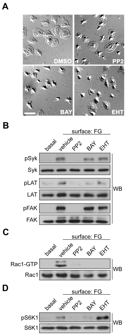
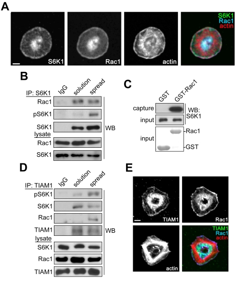
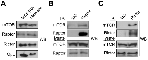
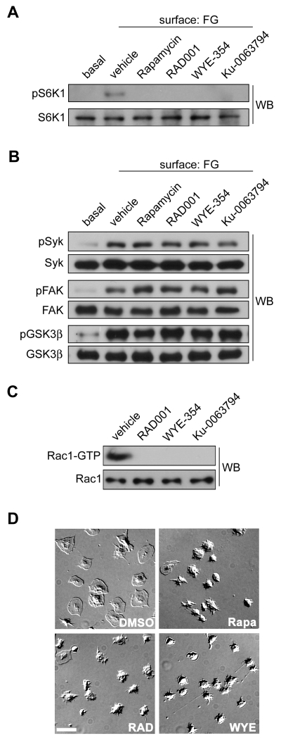
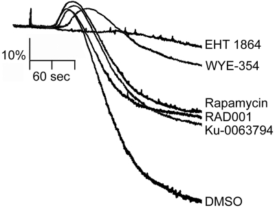
Platelet activation and thrombus formation are under the control of signaling systems that integrate cellular homeostasis with cytoskeletal dynamics. Here, we identify a role for the ribosome protein S6 kinase (S6K1) and its upstream regulator mTOR in the control of platelet activation and aggregate formation under shear flow. Platelet engagement of fibrinogen initiated a signaling cascade that triggered the activation of S6K1 and Rac1. Fibrinogen-induced S6K1 activation was abolished by inhibitors of Src kinases, but not Rac1 inhibitors, demonstrating that S6K1 acts upstream of Rac1. S6K1 and Rac1 interacted in a protein complex with the Rac1 GEF TIAM1 and colocalized with actin at the platelet lamellipodial edge, suggesting that S6K1 and Rac1 work together to drive platelet spreading. Pharmacologic inhibitors of mTOR and S6K1 blocked Rac1 activation and prevented platelet spreading on fibrinogen, but had no effect on Src or FAK kinase activation. mTOR inhibitors dramatically reduced collagen-induced platelet aggregation and promoted the destabilization of platelet aggregates formed under shear flow conditions. Together, these results reveal novel roles for S6K1 and mTOR in the regulation of Rac1 activity and provide insights into the relationship between the pharmacology of the mTOR system and the molecular mechanisms of platelet activation.


Figures

Figure 1
Figure 1
S6K1 is activated upstream of Rac1 in platelets. (A) Representative DIC images of purified human platelets (2 × 10⁷/mL) treated with vehicle (DMSO), the Src inhibitor PP2 (20μM), the Syk inhibitor BAY 61-3606 (1μM) or the Rac1 inhibitor EHT 1864 (50μM), on a surface of fibrinogen (FG). Scale bar = 10 μm. (B) Lysates from quiescent platelets in solution (basal) or FG-surface-attached platelets were analyzed for Src, Syk and FAK activation by Western blotting (WB) for Syk-pTyr323, LAT-pTyr171 and FAK-pTyr576/577. (C) Lysates were incubated with glutathione-sepharose conjugated to GST-PAK-CRIB to capture activated GTP-bound Rac1. Captured GTP-Rac1 and total Rac1 inputs were analyzed by Western blotting. (D) Platelets were treated with inhibitors as above and analyzed for S6K1 activation by Western blotting for S6K1-pThr389. PP2 and BAY 61-3606 decreased S6K1 phosphorylation by 86.9% and 66.9%, respectively (n = 3, P < .05). EHT 1864 increased pS6K1 levels by 121% relative to vehicle (n = 3, P < .05).
Figure 2
Figure 2
S6K1 and Rac1 localize to the lamellipodial edge of spreading platelets. (A) Purified human platelets were spread on coverglass coated with 25 μg/mL fibrinogen. After 45 minutes, platelets were fixed, stained for S6K1, Rac1, and actin, and visualized by confocal microscopy. Scale bar = 2 μm. (B) S6K1 was immunoprecipitated (IP) from lysates of quiescent platelets in solution or platelets spread on fibrinogen and analyzed for coprecipitating Rac1 by Western blot. Nonspecific rabbit immunoglobulins (IgG) were used as negative control for immunoprecipitations. Total S6K1 and Rac1 levels in whole-platelet lysates serve as input controls. (C) Platelet lysates were incubated with GST or Rac1-GST glutathione sepharose and captured S6K1 was analyzed by Western blot. Coomassie-stained GST and Rac1-GST inputs are shown. (D) TIAM1 was immunoprecipitated from platelet lysates as above and examined for coprecipitating S6K1 and Rac1 by Western blot. (E) Localization of TIAM1, Rac1 and actin in fibrinogen-activated human platelets visualized by confocal microscopy. Scale bar = 2 μm. Western blot, IP, protein capture, and imaging results are representative of 3 independent experiments.
Figure 3
Figure 3
mTORC1 and mTORC2 complexes are present in human platelets. (A) Centrifugally cleared lysates-(50 μg) from human platelets or MCF10A breast epithelial cells were analyzed by Western blot for levels of mTOR, Raptor, Rictor, or GβL. (B) mTORC1 complexes were identified by immunoprecipitating Raptor from platelet lysates and Western blotting for associated mTOR. (C) mTORC2 complexes were identified by immunoprecipitation of Rictor followed by Western blotting for coprecipitating mTOR. Nonspecific immunoglobulins (IgG) served as a negative control for immunoprecipitations. Immunoprecipitation results are representative of 3 independent experiments.
Figure 4
Figure 4
mTOR and S6K1 inhibition blocks Rac1 activation and platelet spreading. (A) Purified human platelets were treated with apyrase (2 units/mL) and vehicle (DMSO), Rapamycin (1μM), RAD001 (1 μM), WYE-354 (10μM), or Ku-0063794 (10μM) before spreading on a surface of fibrinogen. Lysates from surface-bound platelets were analyzed for S6K1 activation by Western blotting for S6K1-pThr389. (B) Western blot analysis of Syk-pTyr323, FAK-pTyr576/577, and GSK3β-pSer9 phosphorylation of fibrinogen-activated platelets pretreated with Rapamycin, RAD001, WYE-354, or Ku-0063794. (C) Lysates from surface-bound platelets were incubated with PAK-CRIB-GST to capture activated Rac1. Captured GTP-Rac1 and total Rac1 levels were determined by Western blot. (D) Representative DIC images of human platelets spread on coverglass coated with 100 μg/mL fibrinogen after treatment with apyrase and vehicle (DMSO), Rapamycin, RAD001, or WYE-354. Scale bar = 10 μm. Results are representative of 3 independent experiments.
Figure 5
Figure 5
Inhibitors of mTOR and S6K1 block platelet aggregation. Washed human platelets (2 × 10⁸/mL) were stimulated with 10 μg/mL collagen in the presence of 2 U/mL apyrase and the change in optical density indicative of platelet aggregation was recorded after 10 minutes of preincubation with EHT 1864 (50μM), WYE-354 (10μM), Ku-0063794 (10μM), Rapamycin (1μM), RAD001 (1μM), or vehicle (DMSO). One experiment representative of 3 separate experiments is shown.
Figure 6
Figure 6
mTOR is required for platelet aggregate stability under flow. (A) Purified human platelets (2 × 10⁷/mL) were spread on glass coverslips coated with 100 μg/mL fibrinogen. After 30 minutes, unbound platelets were removed and activated platelets were treated with vehicle (DMSO), WYE-354 (10μM), or EHT 1864 (50μM) for an additional 30 minutes. Platelet spreading and lamellipodial withdrawal were evaluated by DIC microscopy. Scale bar = 10 μm. (B) PPACK-anticoagulated blood was perfused over collagen for 4 minutes to produce platelet aggregates that were subsequently perfused with buffer containing fibrinogen for an additional 4 minutes before perfusion with buffer, fibrinogen, and vehicle (DMSO), WYE-354 (10μM), or EHT 1864 (50μM). Representative DIC images of platelet aggregates are shown. Arrow indicates direction of flow. Scale bar = 50 μm. (C) Quantification of platelet disaggregation promoted by WYE-354 or EHT 1864 under flow (n = 3). Data are represented as mean ± SEM.

Comment in

References

    1. Furie B, Furie BC. Thrombus formation in vivo. J Clin Invest. 2005;115(12):3355–3362. - PMC - PubMed
    1. Ruggeri ZM. Platelets in atherothrombosis. Nat Med. 2002;8(11):1227–1234. - PubMed
    1. Watson SP. Platelet activation by extracellular matrix proteins in haemostasis and thrombosis. Curr Pharm Des. 2009;15(12):1358–1372. - PubMed
    1. McCarty OJ, Larson MK, Auger JM, et al. Rac1 is essential for platelet lamellipodia formation and aggregate stability under flow. J Biol Chem. 2005;280(47):39474–39484. - PMC - PubMed
    1. Watson SP, Auger JM, McCarty OJ, Pearce AC. GPVI and integrin alphaIIb beta3 signaling in platelets. J Thromb Haemost. 2005;3(8):1752–1762. - PubMed

Publication types

MeSH terms

Substances