. 2011 Sep 2;286(35):30662-30669.
doi: 10.1074/jbc.M111.267906. Epub 2011 Jul 11.

A role for prolyl 3-hydroxylase 2 in post-translational modification of fibril-forming collagens

Affiliations

A role for prolyl 3-hydroxylase 2 in post-translational modification of fibril-forming collagens

Russell J Fernandes et al. J Biol Chem. .

Abstract

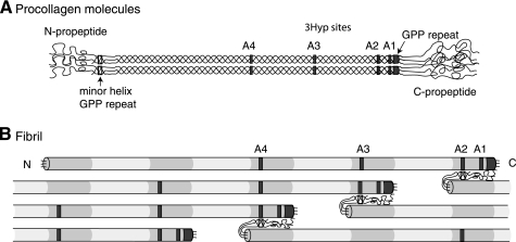
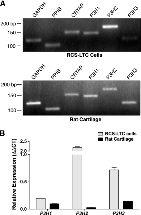
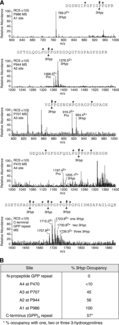
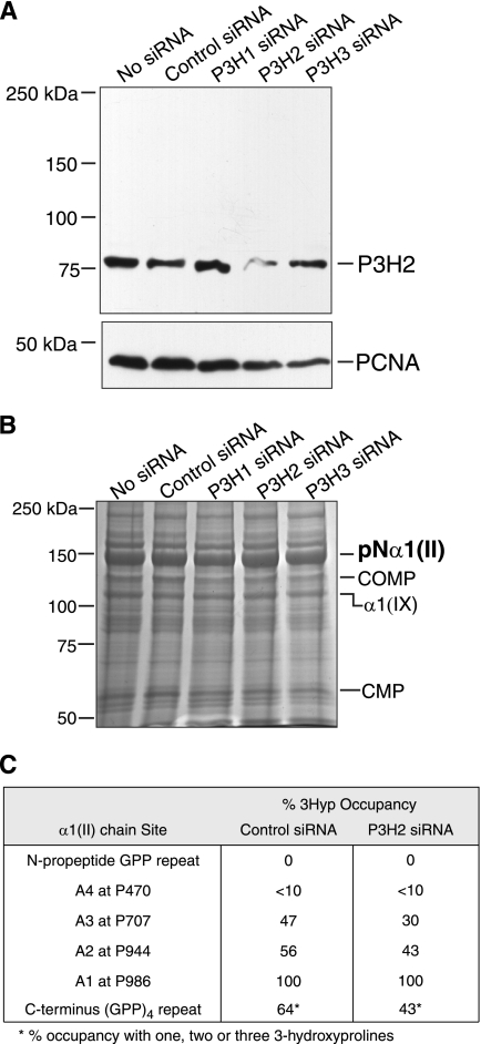
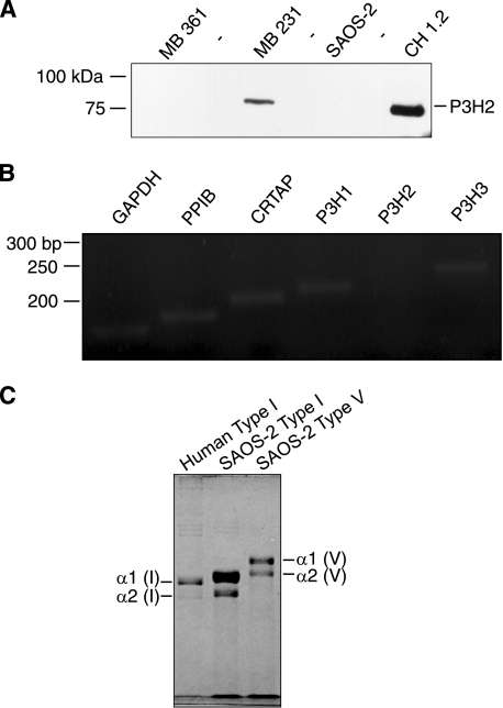
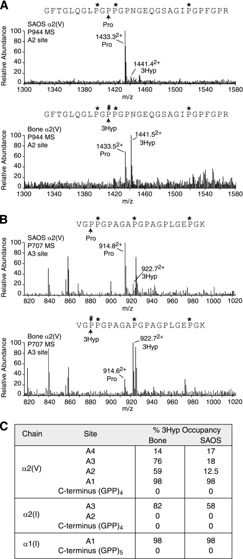
The fibrillar collagen types I, II, and V/XI have recently been shown to have partially 3-hydroxylated proline (3Hyp) residues at sites other than the established primary Pro-986 site in the collagen triple helical domain. These sites showed tissue specificity in degree of hydroxylation and a pattern of D-periodic spacing. This suggested a contributory role in fibril supramolecular assembly. The sites in clade A fibrillar α1(II), α2(V), and α1(I) collagen chains share common features with known prolyl 3-hydroxylase 2 (P3H2) substrate sites in α1(IV) chains implying a role for this enzyme. We pursued this possibility using the Swarm rat chondrosarcoma cell line (RCS-LTC) found to express high levels of P3H2 mRNA. Mass spectrometry determined that all the additional candidate 3Hyp substrate sites in the pN type II collagen made by these cells were highly hydroxylated. In RNA interference experiments, P3H2 protein synthesis was suppressed coordinately with prolyl 3-hydroxylation at Pro-944, Pro-707, and the C-terminal GPP repeat of the pNα1(II) chain, but Pro-986 remained fully hydroxylated. Furthermore, when P3H2 expression was turned off, as seen naturally in cultured SAOS-2 osteosarcoma cells, full 3Hyp occupancy at Pro-986 in α1(I) chains was unaffected, whereas 3-hydroxylation of residue Pro-944 in the α2(V) chain was largely lost, and 3-hydroxylation of Pro-707 in α2(V) and α2(I) chains were sharply reduced. The results imply that P3H2 has preferred substrate sequences among the classes of 3Hyp sites in clade A collagen chains.


Figures

FIGURE 1.
FIGURE 1.
Relative molecular positions of prolyl 3-hydroxylation sites in aligned procollagen molecules and a microfibril. A, occupied 3Hyp sites are indicated by gray boxes within the triple helical regions of the molecule. From right to left, these are the C-terminal (GPP)4 repeat, A1 at Pro-986, A2 at Pro-944, A3 at Pro-707, and A4 at Pro-470. The GPP repeat in the minor triple helix of the N-propeptide is indicated by an open box. B, axial relationships required for trifunctional intermolecular cross-link formation in a pN type II collagen microfibril. Collagen molecules are aligned in the typical collagen D-periodic stagger of 234 amino acid residues. Note the position of the N-propeptide GPP repeat (open box) relative to an A2 site in an adjacent pN type II collagen molecule and alignment of D-staggered A2, A3, and A4 3Hyp sites.
FIGURE 2.
FIGURE 2.
Expression of PPIB, CRTAP, P3H1, P3H2, and P3H3 in the RCS-LTC cell line and rat cartilage. A, RCS-LTC cells transcribe a full complement of the genes assayed. When compared with adult normal rat cartilage, the RCS-LTC cells express an abundance of P3H2. B, quantitative comparison by quantitative PCR of P3H1, P3H2, and P3H3 mRNA expression between RCS-LTC cells and normal adult rat cartilage yielded modestly increased expression of P3H1, an ∼2-fold increase in P3H3 expression and an excess of 8-fold increase in P3H2 in the RCS-LTC cells. Data represents n = 3 for each isoenzyme.
FIGURE 3.
FIGURE 3.
Tandem mass-spectrometric analysis of the RCS-LTC pNα1(II) collagen chain. A, full scan mass spectra from the tryptic peptides LC-MS profiles of pNα1(II) across the elution window of the post-translational variants containing Pro-986, Pro-944 and Pro-707, Pro-470, and the C-terminal (GPP)4 repeat. The relative abundance of the ions shown provides an index of the degree of 3-hydroxylation at the A1, A2, A3, A4, and C-terminal GPP repeat sites. As shown, Pro-944 is 56% 3Hyp occupied and Pro-986 is 100% 3Hyp occupied. B, summary of the relative abundance (%) of 3-hydroxylation of proline residues at sites in pNα1(II) collagen chains from RCS-LTC extracellular matrix.
FIGURE 4.
FIGURE 4.
RNA interference of P3H2 in cultures of the RCS-LTC cell line. A, Western blot of equal aliquots of cell lysates from siRNA-treated cultures probed for P3H2 protein. The P3H2 antibody robustly reacted with an 80-kDa band in siRNA-untreated and control siRNA-treated cultures. A clear reduction of band intensity was specifically observed only in P3H2 siRNA-treated cultures. Proliferating cell nuclear antigen protein was detected as a loading control. Globular protein molecular weight standards were used. B, Coomassie Blue stained gel showing similar amounts of pNα1(II) collagen chains extracted from P3H2 siRNA-treated and untreated cultures. This band from control siRNA and P3H2 siRNA-treated lanes was analyzed by mass spectrometry. Cartilage oligomeric matrix protein (COMP), α1(IX) collagen chain and cartilage matrix protein 1 (CMP) synthesized by the cell line were also identified by mass spectrometry. Globular protein molecular weight standards were run. C, degree (%) of 3-hydroxylation of proline residues in pNα1(II) collagen chains determined by tandem mass spectrometry. The pNα1(II) chains from P3H2 siRNA-treated and control siRNA-treated lanes were subjected to in-gel trypsin digestion, and peptides containing 3Hyp sites were analyzed. 3Hyp occupancy fell at all non-A1 sites on suppression of P3H2 protein.
FIGURE 5.
FIGURE 5.
Expression of PPIB, CRTAP, P3H1, P3H2, and P3H3 in the SAOS-2 cell line. A, cell lysates from MBA 361 and MBA 231 human breast cancer cells, CH1.2 human chondrosarcoma cells, and SAOS-2 human osteosarcoma cells were probed with P3H2 antibody on Western blots. The antibody failed to detect P3H2 protein in the MBA 361 and SAOS-2 cells. B, RT-PCR confirmed the lack of expression of P3H2 by SAOS-2 cells. P3H1, CRTAP, PPIB, and P3H3 were strongly expressed. C, Coomassie Blue-stained gel showing type I collagen (middle lane) and type V collagen (right lane) extracted from the extracellular matrix of cultured SAOS-2 cells. Human bone type I collagen was run as a control (left lane). The α1(I), α2(I), and the α2(V) bands were cut out and analyzed for 3Hyp occupancy by mass spectrometry.
FIGURE 6.
FIGURE 6.
Tandem mass spectrometric analysis of 3Hyp in matrix collagen deposited by SAOS-2 cells. A, B, α2(V) chains from the SAOS-2 cell line were compared with human bone. Full scan mass spectra from the tryptic peptide LC-MS profiles of α2(V) across the elution window of the post-translational variants containing Pro-986 (A1), Pro-944 (A2), and Pro-707 (A3) are shown. C, summary of % prolyl 3-hydroxylation levels at sites in α2(V), α2(I), and α1(I) collagen chains from SAOS-2 cells and normal human bone.

References

    1. Olsen B. R., Berg R. A., Kivirikko K. T., Prockop D. J. (1973) Eur. J. Biochem. 35, 135–147 - PubMed
    1. Risteli J., Kivirikko K. I. (1974) Biochem. J. 144, 115–122 - PMC - PubMed
    1. Kivirikko K. I., Ryhänen L., Anttinen H., Bornstein P., Prockop D. J. (1973) Biochemistry 12, 4966–4971 - PubMed
    1. Myllylä R., Alitalo K., Vaheri A., Kivirikko K. I. (1981) Biochem. J. 196, 683–692 - PMC - PubMed
    1. Vranka J. A., Sakai L. Y., Bächinger H. P. (2004) J. Biol. Chem. 279, 23615–23621 - PubMed

Publication types