. 2011 Sep 23;286(38):33158-66.
doi: 10.1074/jbc.M111.250456. Epub 2011 Jul 13.

Characterization of the deoxynucleotide triphosphate triphosphohydrolase (dNTPase) activity of the EF1143 protein from Enterococcus faecalis and crystal structure of the activator-substrate complex

Affiliations

Characterization of the deoxynucleotide triphosphate triphosphohydrolase (dNTPase) activity of the EF1143 protein from Enterococcus faecalis and crystal structure of the activator-substrate complex

Ivan I Vorontsov et al. J Biol Chem. .

Abstract

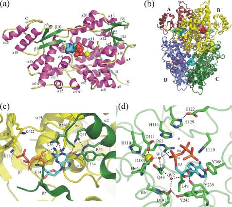
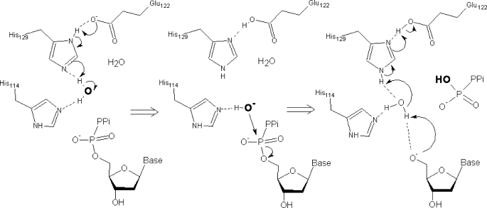
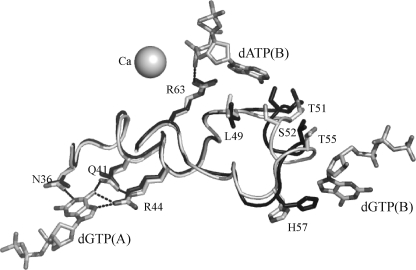
The EF1143 protein from Enterococcus faecalis is a distant homolog of deoxynucleotide triphosphate triphosphohydrolases (dNTPases) from Escherichia coli and Thermus thermophilus. These dNTPases are important components in the regulation of the dNTP pool in bacteria. Biochemical assays of the EF1143 dNTPase activity demonstrated nonspecific hydrolysis of all canonical dNTPs in the presence of Mn(2+). In contrast, with Mg(2+) hydrolysis required the presence of dGTP as an effector, activating the degradation of dATP and dCTP with dGTP also being consumed in the reaction with dATP. The crystal structure of EF1143 and dynamic light scattering measurements in solution revealed a tetrameric oligomer as the most probable biologically active unit. The tetramer contains four dGTP specific allosteric regulatory sites and four active sites. Examination of the active site with the dATP substrate suggests an in-line nucleophilic attack on the α-phosphate center as a possible mechanism of the hydrolysis and two highly conserved residues, His-129 and Glu-122, as an acid-base catalytic dyad. Structural differences between EF1143 apo and holo forms revealed mobility of the α3 helix that can regulate the size of the active site binding pocket and could be stabilized in the open conformation upon formation of the tetramer and dGTP effector binding.


Figures

FIGURE 1.
FIGURE 1.
a, general view of the EF1143 polypeptide chain B from the holo (3IRH) crystal structure. The dATP substrate and Ca2+ ion are shown as spheres. b, EF1143 homotetramer with approximate 222 symmetry. dATP substrates and dGTP effectors are shown as red and cyan spheres, respectively. c, detailed view of the effector binding site in the interface of two polypeptide chains A (green) and B (yellow). One side of the binding pocket is formed by an antiparallel β-sheet (strands β2 and β3) and helices α1, α2, and α4 of one monomer, another, by the secondary structure elements of the adjacent polypeptide chain: helices α3 and α14, and loops α18→β6 and β10→α21. d, active site in chain B(3IRH). A magenta star designates the place where a water molecule could be activated and localized to perform an in-line nucleophilic attack on Pα-O5 bond. His-129–Glu-122 is a putative catalytic dyad. His-114 could assist in correct positioning of the attacking water. A possible function of highly conserved residues Gln-48, His-61, Asp-191, and Tyr-243 would be to provide H-bond network for the structural W2 water molecule which, together with Tyr-239, Arg-63, and W1, appears to play an important role in recognition and proper positioning of the deoxyribose unit of the substrate.
FIGURE 2.
FIGURE 2.
Scheme of a possible mechanism of the hydrolysis.
FIGURE 3.
FIGURE 3.
Mechanism of the allosteric regulation of the EF1143 activity. Superposition of the chain A(2O6I) (black) from the apo structure with the chain B(3IRH) (gray) from the holo form together with Ca2+, two dGTPs and dATP. Formation of the tetramer and binding of the dGTP(B) effector results in the stabilization of the α3 helix, residues 52–56, in the “open” conformation (gray). In the “closed” conformation (black) the Ser-52 residue protrudes its side chain into the interior of the binding pocket, thus, preventing docking of the substrate into the active site.

References

    1. Huber H. E., Beauchamp B. B., Richardson C. C. (1988) J. Biol. Chem. 263, 13549–13556 - PubMed
    1. Aravind L., Koonin E. V. (1998) Trends Biochem. Sci. 23, 469–472 - PubMed
    1. Finn R. D., Tate J., Mistry J., Coggill P. C., Sammut S. J., Hotz H. R., Ceric G., Forslund K., Eddy S. R., Sonnhammer E. L., Bateman A. (2008) Nucleic Acids Res. 36, D281–D288 - PMC - PubMed
    1. Kornberg S. R., Lehman I. R., Bessman M. J., Simms E. S., Kornberg A. (1958) J. Biol. Chem. 233, 159–162 - PubMed
    1. Quirk S., Bessman M. J. (1991) J. Bacteriol. 173, 6665–6669 - PMC - PubMed

Publication types

MeSH terms

LinkOut - more resources