. 2011 Sep 16;286(37):31993-2001.
doi: 10.1074/jbc.M111.219212. Epub 2011 Jul 13.

The T cell receptor triggering apparatus is composed of monovalent or monomeric proteins

Affiliations

The T cell receptor triggering apparatus is composed of monovalent or monomeric proteins

John R James et al. J Biol Chem. .

Abstract

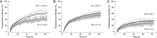
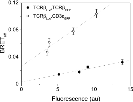
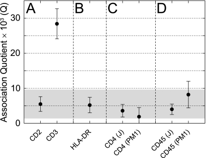
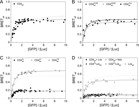
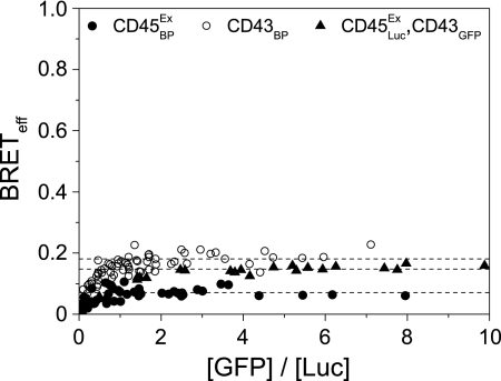
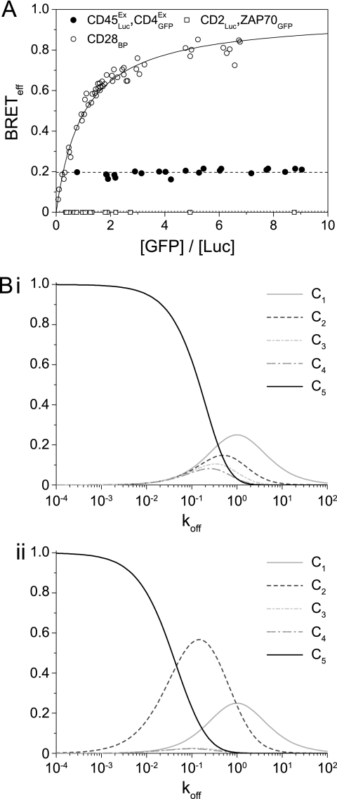
Understanding the component stoichiometry of the T cell antigen receptor (TCR) triggering apparatus is essential for building realistic models of signal initiation. Recent studies suggesting that the TCR and other signaling-associated proteins are preclustered on resting T cells relied on measurements of the behavior of membrane proteins at interfaces with functionalized glass surfaces. Using fluorescence recovery after photobleaching, we show that, compared with the apical surface, the mobility of TCRs is significantly reduced at Jurkat T cell/glass interfaces, in a signaling-sensitive manner. Using two biophysical approaches that mitigate these effects, bioluminescence resonance energy transfer and two-color coincidence detection microscopy, we show that, within the uncertainty of the methods, the membrane components of the TCR triggering apparatus, i.e. the TCR complex, MHC molecules, CD4/Lck and CD45, are exclusively monovalent or monomeric in human T cell lines, implying that TCR triggering depends only on the kinetics of TCR/pMHC interactions. These analyses also showed that constraining proteins to two dimensions at the cell surface greatly enhances random interactions versus those between the membrane and the cytoplasm. Simulations of TCR-pMHC complex formation based on these findings suggest how unclustered TCR triggering-associated proteins might nevertheless be capable of generating complex signaling outputs via the differential recruitment of cytosolic effectors to the cell membrane.


Figures

FIGURE 1.
FIGURE 1.
Nonspecific T cell binding to a glass surface leads to changes in TCR diffusion. FRAP analysis of Alexa Fluor 488-labeled TCR (CD3ϵ) is presented. Mean recovery curves are shown with data bounded by 95% confidence intervals at each point, with the apical surface in solid circles and basal in open circles for Jurkat cells [n = 20] (A), J45 cells [n = 32] (B) and J.CaM1.6 cells [n = 20] (C). The mean ± S.E. of asymptotic fluorescence recovery for each dataset are also shown.
FIGURE 2.
FIGURE 2.
The Jurkat TCR complex contains only one β-chain. Jurkat cells co-expressing TCRβ fused to luciferase or GFP show low energy transfer, as detected by BRET (filled circles). Expression of CD3ϵGFP with TCRβLuc gave significant BRET that does not extrapolate to zero at low acceptor levels, as expected for a specific association (open circles). GFP fluorescence was measured by flow cytometry.
FIGURE 3.
FIGURE 3.
The endogenous membrane proteins of the triggering apparatus are monomeric at the cell surface. A, Fabs against the known monomer CD2 define the expected Q value from TCCD for noninteracting proteins within the membrane. The presence of two CD3ϵ chains per TCR complex is confirmed by TCCD and constitutes the positive control. B, HLA-DR expressed on THP-1 monocyte cells showed no evidence for a dimer of dimers. C, the CD4 co-receptor on two independent T cell lines is monomeric by TCCD. D, CD45 is also shown to be monomeric on both of these cell types. The shaded part of the graph is a 95% confidence interval based on the CD2 dataset that defines the most likely Q values for monomeric proteins.
FIGURE 4.
FIGURE 4.
CD4 co-receptor is monomeric in the presence of Lck kinase. A, expression of CD4 as a BRET pair in HEK-293T cells shows oligomerization (fitted solid line) that does not fit as well to a dimer model (dotted line). B, mutation of residues thought to disrupt CD4 oligomerization or of the intracellular cysteines had a negligible effect on BRETeff values; the fit to wild type CD4 (from A) is shown for comparison in this and all subsequent panels (dotted line). C, truncations of the co-receptor isolated the intracellular region of CD4 as the driving force for oligomerization. D, co-expression of Lck with CD4 leads to the complete and specific disruption of oligomerization to levels equivalent to the monomeric controls and Lck itself.
FIGURE 5.
FIGURE 5.
The extracellular domain of CD45RO is monomeric. The BRET assay was used to determine the level of CD45 self-association in HEK-293T cells. No specific oligomerization of the phosphatase could be detected compared with appropriate controls. All data were fitted as monomers (dashed lines).
FIGURE 6.
FIGURE 6.
Interactions within the membrane are highly favored compared with those with molecules from the cytosol. A, the BRETeff values for a CD45/CD4 pair are nonspecific but significant when compared with those of the covalent homodimer CD28. Co-expression of the monomer CD2 with cytosolic GFP-tagged ZAP70 gives undetectable energy transfer. B, data from the molecular simulation are shown, with the relative proportions of the various complexes [C] with the original (i) or revised (ii) KP model, as the koff for the TCR/pMHC interaction is varied.

Similar articles

Cited by

References

    1. Smith-Garvin J. E., Koretzky G. A., Jordan M. S. (2009) Annu. Rev. Immunol. 27, 591–619 - PMC - PubMed
    1. Salzmann M., Bachmann M. F. (1998) Mol. Immunol. 35, 271–277 - PubMed
    1. Schamel W. W., Arechaga I., Risueño R. M., van Santen H. M., Cabezas P., Risco C., Valpuesta J. M., Alarcón B. (2005) J. Exp. Med. 202, 493–503 - PMC - PubMed
    1. James J. R., White S. S., Clarke R. W., Johansen A. M., Dunne P. D., Sleep D. L., Fitzgerald W. J., Davis S. J., Klenerman D. (2007) Proc. Natl. Acad. Sci. U.S.A. 104, 17662–17667 - PMC - PubMed
    1. Lillemeier B. F., Mörtelmaier M. A., Forstner M. B., Huppa J. B., Groves J. T., Davis M. M. (2010) Nat. Immunol. 11, 90–96 - PMC - PubMed

Publication types

MeSH terms