Review
. 2011 Sep;18(9):1487-99.
doi: 10.1038/cdd.2011.81. Epub 2011 Jul 15.

p63 is a suppressor of tumorigenesis and metastasis interacting with mutant p53

Affiliations
Review

p63 is a suppressor of tumorigenesis and metastasis interacting with mutant p53

G Melino. Cell Death Differ. 2011 Sep.

Abstract

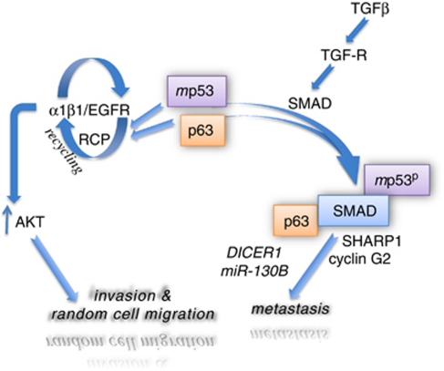
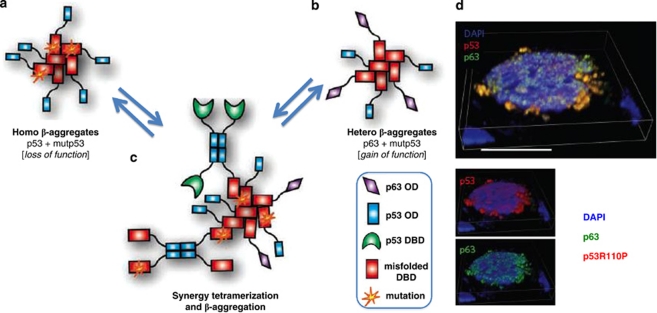
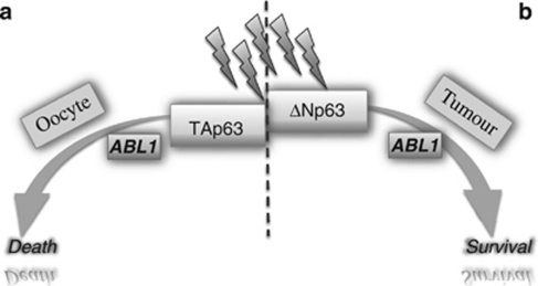
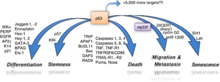
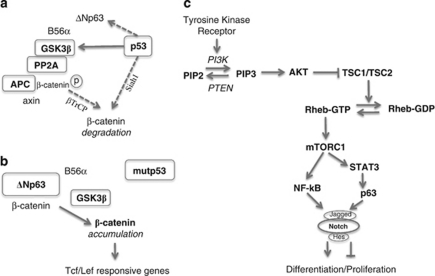
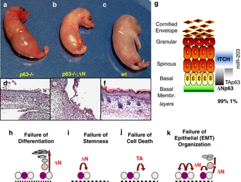
p53 mutations, occurring in two-thirds of all human cancers, confer a gain of function phenotype, including the ability to form metastasis, the determining feature in the prognosis of most human cancer. This effect seems mediated at least partially by its ability to physically interact with p63, thus affecting a cell invasion pathway, and accordingly, p63 is deregulated in human cancers. In addition, p63, as an 'epithelial organizer', directly impinges on epidermal mesenchimal transition, stemness, senescence, cell death and cell cycle arrest, all determinant in cancer, and thus p63 affects chemosensitivity and chemoresistance. This demonstrates an important role for p63 in cancer development and its progression, and the aim of this review is to set this new evidence that links p63 to metastasis within the context of the long conserved other functions of p63.


Figures

Figure 1
Figure 1
The p63 proteins. The TP63 gene (a) codifies several proteins (b) thanks to two distinct promoters (P1 and P2) and 3′ alternative splicing. In addition to the full length α isoform, two isoforms have been described: a β isoform (skipping exon 13) and a γ isoform (alternative exon 15, following exon 10, with its stop codon and distinct 3′-UTR. In silico analysis predicted the δ isoform (skipping exon 12–13) and the ɛ isoform (premature termination in intron 10 retaining the 5′-portion of intron 10 with a stop codon). The structure is currently available only for the DBD (MMDB ID: 67838 PDB ID: 2RMN) (c) and for the SAM domain (MMDB ID: 30268 PDB ID: 1RG6) (d). The principal domains are shown, with their identity with both p73 and p53. TA, transactivation domain (aa 1–64, residues of human TAp63); PR, proline-rich domain; DBD, DNA-binding domain (aa 142–323); OD, oligomerization domain (aa 353–397); TA2, second transactivation domain (aa 410–512); SAM, sterile alpha motif (aa 502–566); TI, transcription inhibitory domain (aa 568–641)
Figure 2
Figure 2
Mutant p53 and p63 as a suppressor of metastasis. Two models showing how mutant p53 gains a novel function through its ability to interact with p63, thus becoming able to affect cell invasion and metastasis. According to REF (left arm) mutant p53 interacts with p63 and regulates integrin recycling in a Rab-coupling protein (RCP)-dependent manner. This increases the intracellular steady-state levels of AKT and thus affects random cell mobility and invasion. According to REF (right arm) transforming growth factor-β (TGFβ) binds its receptor (TGFR), allowing the increase in SMAD2, which is able to bind mutant p53 (mp53) that has been phosphorylated in a NRAS-dependent manner (not shown in the figure). A tripartite complex is then formed, with SMAD2 bridging mutant p53 to p63. This results in the regulation of metastasis through SHARP1 (also known as BHLHE41) and cyclin G2 through integrin endocytosis recycling or through DICER1 and miR-130B,
Figure 3
Figure 3
Interactions and aggregations within the p53 family members. Conformationally destabilized mutant p53 forms aggregate with wild type p53 (a), acting as a dominant-negative (in addition to competing for the same promoter-binding sites), or with p63 (b), causing a gain-of-function effect because of altered p63 transcriptional activity. Interactions through the OD (c), recruits additional p63 in the aggregates, augmenting the effect of mutant p53. (d) Colocalization of p63 (green) with mutant p53 (red) in the nucleus (blue). DBD, DNA-binding domain; OD, oligomerization domain. Modified with permission from Xu et al.
Figure 4
Figure 4
Phosphorylation of p63 regulates death and survival in cancer cells and in oocytes. The TAp63 isoform seems to be specific for oocytes (a) while ΔNp63 is highly expressed in cancer cells (b). Both isoforms require phosphorylation on specific residues by the ABL kinase to become activated,, and, following DNA damage, induce death of oocytes and survival of cancer cells. Consequently, ABL inhibition could affect the survival of oocytes (left), while disrupting a relevant survival mechanism in cancer cells (right)
Figure 5
Figure 5
Cancer pathways involving p63. p63 can potentially transactivate several thousand target genes, regulating several biochemical pathways that are relevant to tumorigenesis. Although p63 can transactivate the same targets as p53, indicating a high similarity in the interaction with the responsive element, p63 has indeed distinct targets; as all p63 isoforms share the same DNA-binding domain, there is no significant qualitative difference in the recognition of the responsive element but the composition of the transcription complex seems to be distinct, resulting in different biological effects. First, TAp63 is able to regulate cell death through several distinct mechanisms, depending on the stressor and the tissue involved (death). Second, ΔNp63 controls the formation of the epidermis,, and thus it is implicated in related tumors, acting on specific promoters, some of which are illustrated schematically (differentiation). However, a crucial role in cancer seems to be on the proliferation potential of the stem cell compartment, although in this case, the underlying molecular targets are still not fully clear (stemness). This activity strongly suggests a corresponding role in cancer stem cells, though this has yet to be formally proven. Furthermore, p63 regulates the senescence pathway both in normal and in cancer cells,, (senescence), via distinct yet unclear targets. Finally, p63, neutralized in cancer cells by binding to mutant p53, regulates an adhesion and epithelial–mesenchymal transition pathway that is crucial for migration, invasion and metastasis, , (migration and metastasis). The main transcriptional targets directly regulated by p63 are indicated in their current nomenclature
Figure 6
Figure 6
p63 signaling in cancer cells. Opposing effects of ΔNp63 and β-catenin in cancer cells. In normal cells (a), p53 facilitates the degradation of ΔNp63 and seems to activate GSK3β, causing degradation of β-catenin. In squamous cell lung cancer (b), mutant p53 fails to downregulate ΔNp63, which binds B56α, inhibiting GSKβ and altering the β-catenin APC-binding complex, with accumulation of non-phosphorylated β-catenin. (c) STAT3/p63 regulate Notch signaling downstream of TORC1 to affect the molecular switch of differentiation versus proliferation. NF-kB counteracts this pathway
Figure 7
Figure 7
Potential functions for p63 in the epidermis. Trp63−/− mice have no skin or limbs (a) as compared with wt (c), because of the total absence of epidermis (d versus f). This is mainly due to the ΔNp63 isoform, as seen from genetic complementation studies (b and e). In this study, for example, the p63-null mice were crossed with a transgenic mouse overexpressing TAp63 or ΔNp63 under the control of the promoter of keratin 5, in the basal layer; complementation with ΔNp63 (b and e), but not with TAp63, partially rescued the phenotype indicating that ΔNp63 is the isoform predominantly responsible for the absence of epidermis in the full knockout mice (a and d). p63 is expressed in the basal layer of the epidermis (g) as the ΔNp63 isoform (99% by PCR in adult mice), and its localization is restricted to the basal layer by the action of the E3 ubiquitin ligase ITCH as well as by miR-203. Modified with permission from. The major role of ΔNp63 during development is, therefore, in the formation of the epidermis through a mechanism not fully clarified at the molecular level. This is important as it could also highlight the potential role of p63 in cancer (Figures 5 and 6). Distinct hypotheses were initially formulated, assigning a crucial role for ΔNp63 in ‘differentiation' (h), for example, transactivating K14, IKKα, of pluri-stratified epithelia. Recent data unveiled the role of ΔNp63 in regulating the proliferation potential of the ‘stem cell' (i) compartment, although the underlying molecular mechanisms have not been fully clarified yet. In addition to being essential in development, this pathway could also be important in cancer. On the other hand, TAp63 seems to be mainly involved in ‘cell death' (j), for example, transactivating Puma, Noxa, CD95, Bax. This, clearly, could be relevant in adult tumorigenesis. TAp63-related cell death seemed to be mainly involved in defense from exogenous stresses (sun burn cells following UV irradiation), while ΔNp63 seemed to affect the physiological differentiation pathway. Finally, very recent data suggest the existence of a further additional pathway, induced by ΔNp63, able to regulate adhesion and EMT as ‘epithelial organizer' (k), see main text

Similar articles

Cited by

References

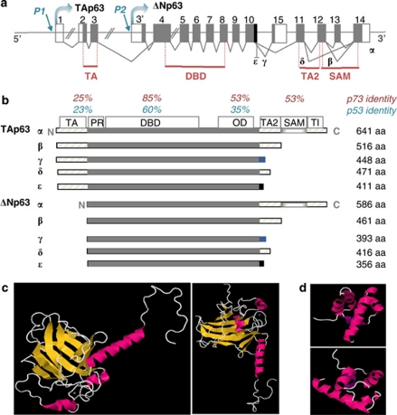
    1. Yang A, Kaghad M, Wang Y, Gillett E, Fleming MD, Dotsch V, et al. p63, a p53 homolog at 3q27-29, encodes multiple products with transactivating, death-inducing, and dominant-negative activities. Mol Cell. 1998;2:305–316. - PubMed
    1. Schmale H, Bambergeric C. A novel protein with strong homology to the tumor suppressor p53. Oncogene. 1997;15:1363–1366. - PubMed
    1. Trink B, Okami K, Wu L, Spiuranpong V, Jen J, Sidransky D. A new human p53 homolog. Nat Med. 1998;4:747–748. - PubMed
    1. Senoo M, Seiki N, Ohira M, Sugano S, Watanabe M, Tachibana M, et al. A second p53-related protein, p73L, with high homology to p73. Biochem Biophys Res Commun. 1998;248:603–607. - PubMed
    1. Senoo M, Seiki N, Ohira M, Sugano S, Watanabe M, Tachibana M, et al. A second p53-related protein, p73L, with high homology to p73. Biochem Biophys Res Commun. 1998;248:603–607. - PubMed

Publication types

MeSH terms