

Review
doi: 10.1038/nri3020.
Interrogating the repertoire: broadening the scope of peptide-MHC multimer analysis
Affiliations
- PMID: 21760610
- PMCID: PMC3699324
- DOI: 10.1038/nri3020
Item in Clipboard
Review
Interrogating the repertoire: broadening the scope of peptide-MHC multimer analysis
Nat Rev Immunol.
.
Abstract
Labelling antigen-specific T cells with peptide-MHC multimers has provided an invaluable way to monitor T cell-mediated immune responses. A number of recent developments in this technology have made these multimers much easier to make and use in large numbers. Furthermore, enrichment techniques have provided a greatly increased sensitivity that allows the analysis of the naive T cell repertoire directly. Thus, we can expect a flood of new information to emerge in the coming years.
Conflict of interest statement
The authors declare competing financial interests: see Web version for details.
Figures
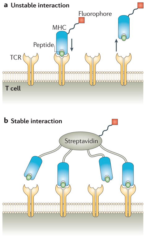
a | As T cell receptors (TCRs) typically dissociate quickly from peptide–MHC complexes (with half-lives of a few seconds), fluorescently labelled monomeric peptide–MHC molecules do not normally survive the washing step during a staining procedure. b | By contrast, if two or more ligands that are part of a tetramer bind simultaneously, then even when one dissociates, others keep the tetramer bound to the cell.

Biotinylated MHC molecules are produced in complex with an ultraviolet (UV)-sensitive MHC-binding peptide. After UV-mediated cleavage of the photolabile peptide with 350 nm light, which exerts no damage on the rest of the protein, empty MHC molecules will readily bind to a ‘rescue peptide’. The MHC molecule bound to this rescue peptide can then be used for peptide–MHC tetramer staining. This UV-mediated peptide exchange reaction can be done on a small scale to allow for the production of a large number of different peptide–MHC tetramers with ease. Image is modified, with permision, from REF. © (2006) Macmillan Publishers Ltd. All rights reserved.

We tested how many different fluorophore-conjugated tetramers that had been loaded with the same peptide could stain the same cells and found that up to six commercially available fluorophore-labelled streptavidin reagents could be used simultaneously. Omitting any number of these six fluorophores can be done in 63 (2 – 1) different ways, so these combinations of fluorophores can, in theory, be used to stain just as many different T cell specificities when all the tetramers are applied to the cells simultaneously. Part a illustrates the simplest version of this concept, whereby three T cell receptor (TCR) specificities can be detected using two colours. That is, cells specific for epitope 1 will be stained only with green (fluorescein isothiocyanate (FITC)-labelled) peptide–MHC tetramers, cells specific for epitope 2 will be stained only with red (phycoerythrin (PE)-labelled) peptide–MHC tetramers and cells specific for epitope 3 will stained by both red and green (FITC- and PE-labelled) peptide–MHC tetramers. Part b shows how this concept can be expanded to identify seven different epitopes using three colours (FITC-, PE- and allophycocyanin (APC)-labelled tetramers).

a | After staining a sample of CD8+ T cells with peptide–MHC tetramers (composed of cytomegalovirus (CMV)–HLA-A2 in this example) in the usual way, the cells are stained with a fluorophore-specific antibody coupled to magnetic iron particles. b | The cells are then passed through a magnetized column, in which the iron-labelled cells are trapped by the magnetic field, whereas most of the unlabelled cells are washed away. c | The tetramer-bound cells are eluted by removing the column from the magnetic field.

Overview of peptide–MHC-related technological developments CLIP, class II-associated invariant chain peptide; TCR, T cell receptor.
References
-
- Weitkamp JH, et al. Generation of recombinant human monoclonal antibodies to rotavirus from single antigen-specific B cells selected with fluorescent virus-like particles. J Immunol Methods. 2003;275:223–237. - PubMed
-
- Doucett VP, et al. Enumeration and characterization of virus-specific B cells by multicolor flow cytometry. J Immunol Methods. 2005;303:40–52. - PubMed
-
- McHeyzer-Williams LJ, McHeyzer-Williams MG. Analysis of antigen-specific B-cell memory directly ex vivo. Methods Mol Biol. 2004;271:173–188. - PubMed
-
- Matsui K, et al. Low affinity interaction of peptide–HC complexes with T cell receptors. Science. 1991;254:1788–1791. - PubMed
Publication types
MeSH terms
Substances
Grants and funding
LinkOut - more resources
Full Text Sources
Other Literature Sources
Research Materials
