. 2011 Sep 1;12(9):931-7.
doi: 10.1038/embor.2011.129.

The death receptor CD95 activates the cofilin pathway to stimulate tumour cell invasion

Affiliations

The death receptor CD95 activates the cofilin pathway to stimulate tumour cell invasion

Ernst J A Steller et al. EMBO Rep. .

Abstract

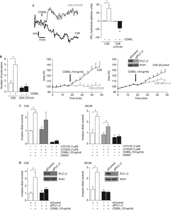
The death receptor CD95 promotes apoptosis through well-defined signalling pathways. In colorectal cancer cells, CD95 primarily stimulates migration and invasion through pathways that are incompletely understood. Here, we identify a new CD95-activated tyrosine kinase pathway that is essential for CD95-stimulated tumour cell invasion. We show that CD95 promotes Tyr 783 phosphorylation of phospholipase C-γ1 through the platelet-derived growth factor receptor-β, resulting in ligand-stimulated phosphatidylinositol (4,5)-bisphosphate (PIP(2)) hydrolysis. PIP(2) hydrolysis liberates the actin-severing protein cofilin from the plasma membrane to initiate cortical actin remodelling. Cofilin activation is required for CD95-stimulated formation of membrane protrusions and increased tumour cell invasion.


Conflict of interest statement

The authors declare that they have no conflict of interest.

Figures

Figure 1
Figure 1
CD95 ligand stimulates rapid cofilin-dependent formation of cell protrusions and an increase in cell area. (A) C26 and C26 KRAS KD cells treated with CD95L (10 ng/ml) for 24 h and stained with propidium iodide. The apoptotic sub-G1 fraction was determined by FACS analysis (left panel). C26 and C26 KRAS KD cells were plated on matrigel-coated membranes in transwell chambers and were allowed to invade for 8 h in the presence or absence of CD95L (10 ng/ml; right panel). The bar graph shows fold increase relative to non-stimulated controls (=1). Error bars represent s.e.m. on the basis of two independent experiments conducted in triplicate. (B) Checkerboard analysis in which CD95L (8 h) was added to the insert (top) or to the well (bottom) of the matrigel invasion chamber. All conditions were tested in three to five independent experiments. Numbers represent fold change of several invaded cells relative to control (no CD95L in either compartment), ±s.e.m. (P-value). Significance was tested using Student's t-test (unpaired; double sided). (C) C26 cells were transiently transfected with LIMK–GFP or GFP and were analysed by real-time imaging before and after stimulation with CD95L (10 ng/ml). Differential interference contrast images were used to determine several protrusions before and after stimulation. Inset shows C26 cells before (left panel) and after (middle panel) CD95L stimulation. The right image is a merge of binary threshold images at both time points. Yellow represents no change; red represents increased protrusion formation; and green represents membrane retraction. Numbers indicate new protrusions formed. Scale bar, 10 μm. The generated images were also used to measure the total cell areas before and after stimulation (arrow) by using ImageJ software (n=4). These values were plotted as the percentage of average cell area before stimulation (right panel). Asterisk indicates significance on the basis of P-values <0.05. (D) C26 and C26–LIMK1 cells were treated with CD95L (10 ng/ml) for 24 h and apoptosis and invasion were assessed as in A (n=3). Asterisk indicates significance on the basis of P-values <0.05. CD95L, CD95 ligand; FACS, fluorescence-activated cell sorting; GFP, green fluorescent protein; KD, knockdown; LIMK, LIM kinase.
Figure 2
Figure 2
CD95 ligand stimulates phospholipase C-γ1 Tyr 783 phosphorylation and PIP2 hydrolysis. (A) C26 cells were treated with CD95L (10 ng/ml) for the indicated time periods. Cell lysates were prepared and analysed for cofilin Ser 3 phosphorylation (pS3–Cofilin), total cofilin, Tyr 783 phosphorylation of PLC-γ1 (pY783–PLC-γ1) and total PLC-γ1 by western blotting. (B) Cells were transiently transfected with mRFP–PH and analysed by real-time confocal microscopy. Images represent mRFP–PH in C26 cells before stimulation (left) and 15 min after stimulation with CD95L (10 ng/ml; middle). Scale bar, 5 μm. Plotting of total cell area (measured by differential interference contrast images) and PIP2 hydrolysis (measured using mRFP–PH) over time shows that addition of CD95L (10 ng/ml; black bar) caused a simultaneous increase in cell area and PIP2 hydrolysis (lower left panel). The average of five independent experiments is plotted (n=5). The right lower panel shows a quantification of maximal CD95L-induced PIP2 hydrolysis, relative to ionomycin-induced maximum translocation of the mRFP–PH probe (100%). Significance was tested using Student's t-test (unpaired; double sided); asterisk indicates P<0.05. CD95L, CD95 ligand; mRFP–PH, PH domain fused to red fluorescent protein; PIP2, phosphatidylinositol (4,5)-bisphosphate; PLC-γ1, phospholipase C-γ1.
Figure 3
Figure 3
CD95 ligand-induced tumour cell invasion requires phospholipase C-γ1 activation. (A) C26 cells were transiently transfected with mRFP–PH. After 24 h, cells were either pretreated with U73122 (5 μM) or with vehicle (DMSO) for 1 h before stimulation with CD95L (10 ng/ml). PIP2 hydrolysis was measured over time, as shown in Fig 2B. (B) C26 cells were either pretreated with U73122 (5 μM) or with vehicle (DMSO) for 1 h before stimulation with CD95L (10 ng/ml). Knockdown of PLC-γ1 was established by transfection of siRNAs 2 days before stimulation. Cells were analysed by real-time imaging. Several cell protrusions and the increase in cell area were measured as shown in Fig 1B. (C) Transwell invasion assays of C26 and MC38 cells treated with U73343 or U73122 were performed as in Fig 1A. Data are from two independent experiments conducted in triplicate. (D) Transwell invasion assays of C26 and MC38 cells transfected with control or PLC-γ1 siRNAs (n=2). Significance was tested using Student's t-test (unpaired; double-sided); asterisk indicates P<0.05. CD95L, CD95 ligand; DMSO, dimethyl sulphoxide; mRFP–PH, PH domain fused to red fluorescent protein; PIP2, phosphatidylinositol (4,5)-bisphosphate; PLC-γ1, phospholipase C-γ1; siRNA, small-interfering RNA.
Figure 4
Figure 4
Platelet-derived growth factor receptor-β mediates CD95-stimulated phospholipase C-γ1 phosphorylation and tumour cell invasion. (A) C26 cells were stimulated with PDGF or EGF and pY783–PLC-γ1 was analysed by western blotting. (B) C26 cells were stimulated with CD95L and PDGFR-β phosphorylation on Tyr 1021 was assessed by western blotting. (C) C26 and MC38 cells were pretreated with sunitinib (2 μM) or AG1296 (20 μM) for 16 h and subsequently stimulated with CD95L for 5, 15 or 30 min. Cell lysates were prepared and analysed for pY783–PLC-γ1 by western blotting. (D) C26 cells were transfected with siRNAs against PDGFR-β and stimulated for the indicated times with CD95L. Y783–PLC-γ1 phosphorylation and PDGFR-β knockdown were analysed by western blotting. Cell area enlargement (right panel) was measured as shown in Fig 1A. (E) Transwell invasion assays of C26 and MC38 cells treated with sunitinib, AG1296 or vehicle (DMSO). Data are from two independent experiments (n=2) conducted in triplicate. (F) Transwell invasion assay of C26 and MC38 cells transfected with control or PDGFR-β-targeting siRNAs (n=2). Significance was tested using Student's t-test (unpaired; double sided), asterisk indicates P<0.05. EGF, epidermal growth factor; PDGF, platelet-derived growth factor; PDGFR-β, PDGF receptor-β; PLC-γ1, phospholipase C-γ1; siRNA, small-interfering RNA.

Similar articles

Cited by

References

    1. Andrianantoandro E, Pollard TD (2006) Mechanism of actin filament turnover by severing and nucleation at different concentrations of ADF/cofilin. Mol Cell 24: 13–23 - PubMed
    1. Arber S, Barbayannis FA, Hanser H, Schneider C, Stanyon CA, Bernard O, Caroni P (1998) Regulation of actin dynamics through phosphorylation of cofilin by LIM-kinase. Nature 393: 805–809 - PubMed
    1. Bamburg JR (1999) Proteins of the ADF/cofilin family: essential regulators of actin dynamics. Annu Rev Cell Dev Biol 15: 185–230 - PubMed
    1. Barnhart BC, Legembre P, Pietras E, Bubici C, Franzoso G, Peter ME (2004) CD95 ligand induces motility and invasiveness of apoptosis-resistant tumor cells. EMBO J 23: 3175–3185 - PMC - PubMed
    1. Bernard O (2007) Lim kinases, regulators of actin dynamics. Int J Biochem Cell Biol 39: 1071–1076 - PubMed

Publication types