Review
. 2011 Oct;25(10):1262-76.
doi: 10.1038/eye.2011.167. Epub 2011 Jul 15.

Adjustable suture strabismus surgery

Affiliations
Review

Adjustable suture strabismus surgery

B R Nihalani et al. Eye (Lond). 2011 Oct.

Abstract

Surgical management of strabismus remains a challenge because surgical success rates, short-term and long-term, are not ideal. Adjustable suture strabismus surgery has been available for decades as a tool to potentially enhance the surgical outcomes. Intellectually, it seems logical that having a second chance to improve the outcome of a strabismus procedure should increase the overall success rate and reduce the reoperation rate. Yet, adjustable suture surgery has not gained universal acceptance, partly because Level 1 evidence of its advantages is lacking, and partly because the learning curve for accurate decision making during suture adjustment may span a decade or more. In this review we describe the indications, techniques, and published results of adjustable suture surgery. We will discuss the option of 'no adjustment' in cases with satisfactory alignment with emphasis on recent advances allowing for delayed adjustment. The use of adjustable sutures in special circumstances will also be reviewed. Consistently improved outcomes in the adjustable arm of nearly all retrospective studies support the advantage of the adjustable option, and strabismus surgeons are advised to become facile in the application of this approach.


Figures
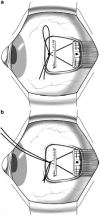
Figure 1
Figure 1
Basic adjustable suture techniques. (a) Bow tie technique: the sutures are tied together in a single-loop bow tie similar to a shoelace. (b) Sliding noose technique: a noose is created by tying a separate piece of suture around the scleral sutures. Reproduced with permission from Hunter et al.
Figure 2
Figure 2
Short tag noose technique showing the fornix incision (F), trimmed pole sutures (P), and trimmed noose (N) buried under the conjunctiva. Reproduced with permission from Hunter et al.
Figure 3
Figure 3
Surgical technique of adjustable SO suture spacer. (a) Exposure of the SO tendon and placement of sutures. (b) Sutures tied, leaving a gap of 2–8 mm. (c) The slipknot is converted into a square knot by cutting one of the pairs of sutures and passing this pair through the knot. The sutures are then secured with a permanent square knot. (d) Final position of SO tendon, with suture spacer in place. Reproduced with permission from Suh et al.
Figure 4
Figure 4
(a) Insertion of SO tendon is exposed lateral to superior rectus (SR) muscle. SO tendon is split about 10 mm back, and suture is sewn through anterior fibers of tendon just behind insertion. (b) Anterior fibers of SO tendon have been disinserted and sutures sewn through partial thickness sclera in more anterior and lateral position. Postoperatively, suture can be allowed to slide back or be pulled forward (see inset boxes) to either weaken or strengthen effect of partial SO tendon advancement. Reproduced with permission from Metz and Lerner.
Figure 5
Figure 5
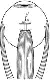
(a) Lateral rectus posterior fixation: 3 mm scleral bite (arrow) taken 18 mm from original insertion. Each arm of a double-armed Mersilene suture is passed through the muscle belly, one-third muscle width from superior and inferior edge (double arrow). (b) The Mersilene suture is then tied over the middle-third of the muscle (arrow). *Indicates the original insertion of the lateral rectus muscle; **indicates lateral rectus muscle. This case shows a simultaneous recession of 7 mm on an adjustable suture. Reproduced with permission from Holmes et al.
Figure 6
Figure 6
Semiadjustable sutures showing that the corners of IR muscle are sutured firmly to the sclera and the center of the muscle is placed on an adjustable suture. Reproduced with permission from Kushner.
Figure 7
Figure 7
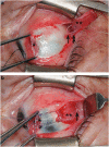
(a) Surgeon's view of the IR muscle. Note the capsulopalpebral head (CPH) of the IR (*) arising from the muscle 4.0–5.0 mm from its insertion. (b) Appearance of the CPH (*) and capsule of the IR muscle following dissection of check ligaments and other fibrous attachments from the muscle. (c) Double-armed 6–0 polyglactin 910 sutures on the IR muscle and on the cut end of the CPH of the muscle. (d) Adjustable suspension of the lower eyelid retractors from the original IR insertion temporal to the muscle's adjustable suture. Reproduced with permission from Pacheco et al.

Comment in

  • Paediatric adjustable strabismus surgery.
    Mokashi A, Stead RE, Stokes J. Mokashi A, et al. Eye (Lond). 2012 Jul;26(7):1024-5; author reply 1025-6. doi: 10.1038/eye.2012.64. Epub 2012 Apr 13. Eye (Lond). 2012. PMID: 22498799 Free PMC article. No abstract available.

References

    1. Bielschowsky A.Die neueren Anschauungen über Wesen und Behandlung des Schielens Med Klin 1907iii335–336.translation by Catharina Latz, MD.
    1. Jampolsky A. Strabismus reoperation techniques. Trans Sect Ophthalmol Am Acad Ophthalmol Otolaryngol. 1975;79:704–717. - PubMed
    1. Jampolsky A. Current techniques of adjustable strabismus surgery. Am J Ophthalmol. 1979;88:406–418. - PubMed
    1. Rosenbaum AL, Metz HS, Carlson M, Jampolsky AJ. Adjustable rectus muscle recession surgery: a followup study. Arch Ophthalmol. 1977;95:817–820. - PubMed
    1. Keech RV, Scott WE, Christensen LE. Adjustable suture strabismus surgery. J Pediatr Ophthalmol Strabismus. 1987;24:97–102. - PubMed

Publication types