. 2011:2011:965016.
doi: 10.1155/2011/965016. Epub 2011 Jul 2.

Seed Oil of Brucea javanica Induces Apoptotic Death of Acute Myeloid Leukemia Cells via Both the Death Receptors and the Mitochondrial-Related Pathways

Affiliations

Seed Oil of Brucea javanica Induces Apoptotic Death of Acute Myeloid Leukemia Cells via Both the Death Receptors and the Mitochondrial-Related Pathways

Hong Zhang et al. Evid Based Complement Alternat Med. 2011.

Abstract

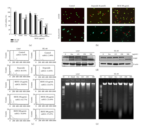
Seed oil of Brucea javanica (BJO) is extracted from the seeds of herb medicine Brucea javanica (L.), and its emulsion formulation (BJOE) has been used clinically to treat carcinomas for many years in China. The antileukemia potential of BJO was investigated in human acute myeloid leukemia cell lines (AML) U937 and HL-60 in vitro and in a mouse U937 xenograft tumor model. BJO induced AML cell apoptosis through activation of caspase-8 and modulation of apoptosis-related proteins. Meanwhile, the inhibition of survivin and XIAP increased the cytotoxicity of BJO. Consistent with these findings, BJO also increased subG(1) phase cells and cause PARP cleavage in AML patients' leukemia cells. In contrast, only weak cytotoxicity of BJO was found in peripheral blood lymphocytes (PBLs) of healthy volunteers. Moreover, oleic acid and linoleic acid were found to be the active components of BJO. Our study provided strong evidence for the first time that BJO induced apoptosis of both cultured and primary AML cells. Furthermore, intravenous injection of BJO significantly inhibited U937 tumor growth in the xenograft mouse model. These results suggest that BJO may have a therapeutic role in the treatment of human leukemia.


Figures

Figure 1
Figure 1
Cytotoxic effects of BJO on different tumor cell lines. HL-60 and U937 cells were treated with emulsifier (negative control), etoposide (10 μmol/L, positive control), or various concentrations of BJO as indicated for 6 hours. (a) Cell viabilities were determined by MTT assay. ***P < .001 (b) U937 cell morphology analysis following treatment with BJO. AO/EB and Hochest3325 were used to stain cells and the cellular fluorescent changes were observed using fluorescence microscope. Results were obtained from three separate experiments. (c) Apoptotic cells were evaluated by cell cycle analysis. Percentage of cells in the subG1 phase was measured using flow cytometry as described in Materials and Method. (d) Western blotting analysis of PARP. β-actin is shown as a loading control. (e) DNA fragmentation analysis. The level of DNA fragmentation was determined as described in Materials and Method.
Figure 2
Figure 2
Effect of BJO on ROS release and mitochondrial membrane potential (MMP) collapse in U937 cells. U937 cells were treated with etoposide (10 μmol/L) or various concentrations of BJO as indicated for 6 h. (a) U937 cells were labeled with 5 μmol/L DCFH-DA for 1 h prior to the treatment with BJO. The addition of 100 μmol/L H2O2 for 1 h was used as a positive control for the H2O2 level. (b) Disruption of MMP was determined according to changes in fluorescence density using rhodamine 123. (c, d, and e). ROS accumulation, MMP, and cell cycle examination of U937 pretreated with antioxidants. Cells were pretreated with catalase (500 U/mL) or N-acetylcysteine (10 mM) for 4 hours, followed by treatment with or without BJO for another 6 h. ROS level, MMP, and cell cycle were measured using flow cytometry as described in Materials and Methods.
Figure 3
Figure 3
Effect of BJO on apoptotic proteins. AML cells were treated with etoposide (10 μmol/L) and various concentrations of BJO as indicated for 6 hours. (a) The mRNA levels of apoptosis-related proteins were examined by RT-PCR as described in Materials and Methods. (b, c, and d) Western blotting analysis of procaspase-3, procaspase-9, Bcl-1, Mcl-1, Bax, Bid, pro-caspase-8, c-FLIP(L/S), DR4, pro-DR5, Fas, XIAP and Survivin. Lane 1 and Lanes 3–5: cells were treated with or without BJO. Lane 2: cells were treated with etoposide (10 μmol/L). The level of each protein was determined using specific antibodies as described in Materials and Method.
Figure 4
Figure 4
Western blotting analysis of PARP in primary leukemia cells from AML patients. The cells were treated with or without BJO. β-actin was also detected as the loading control. Data were represented as mean ± SE of three separate experiments, ***P < .001. The data was a representative of one result of fifteen patients.
Figure 5
Figure 5
Effect of BJO on human peripheral blood lymphocytes and AML cells. AML cells and PBLs were treated with emulsifier (negative control), etoposide (10 μmol/L, positive control), or indicated concentrations of BJO for 6 hours. (a) Cell viabilities were determined by MTT assay. ***P < .001 (b) Apoptotic cells were evaluated by cell cycle analysis. Percentage of cells in the subG1 phase was measured using FACS as described in Materials and Method. (c) Disruptions of MMP were determined by changes in fluorescence density using rhodamine 123.
Figure 6
Figure 6
BJO inhibits the growth of mouse tumors in U937 xenograft model. (a) U937 cells (3 × 10⁷/200 μL PBS per mouse) were injected s.c. into the right flank of mice. Mice were injected with saline or designated doses of BJO as described in Materials and Methods. Tumor size was measured once every two days with a caliper (calculated volume = shortest diameter2× longest diameter/2). (b) 11th day after intravenously injection BJO, mice were sacrificed and the tumors were excised and weighed. ***P < .001. (c) The picture of tumor in nude mice with or without treatment of BJO.
Figure 7
Figure 7
Total contents of Brucea Javanica oil emulsion. The content of BJOE was analyzed by gas chromatography-mass spectrometry as described in Materials and Methods. The percentages of six components in BJOE were phenol (a; C6H6O, 0.59%), hexadecanoic acid (b; C17H34O2, 11.75%), octadecanoic acid (c; C19H38O2, 5.45%), 9-octadecenoic acid (d; C19H36O2, 29.24%), 9,12-octadecadienoic acid (e; C19H34O2, 44.85%), and 9,12,15-octadecatrienoic acid (f; C19H32O2, 4.22%). Representative graph of two runs.
Figure 8
Figure 8
Active contents of Brucea Javanica oil. (a) HL-60 and U937 cells were treated with oleic acid (37.5, 75, and 150 μg/mL) as indicated for 6 h. (b) HL-60 and U937 cells were treated with linoleic acid (56.25, 112.5, 225 μg/mL) as indicated for 6 h. (c) HL-60 and U937 cells were treated with the mixture of oleic acid and linoleic acid as the the same ratio as in BJO (low:oleic acid 37.5 μg/mL & linoleic acid 56.25 μg/mL; Middle: oleic acid 75 μg/mL & linoleic acid 112.5 μg/mL; High: oleic acid 150 μg/mL & linoleic acid 225 μg/mL) for 6 h. (d) U937 and HL-60 cells morphology analysis following treatment with the mixture of oleic acid and linoleic acid. AO/EB was used to stain cells and the cellular fluorescent changes were observed using fluorescence microscope. Results were obtained from three separate experiments.
Figure 9
Figure 9
A schematic illustration of signal pathways involved BJO-induced cell apoptosis.

References

    1. Su SY. Treatment of lung cancer with brain metastasis using an intravenous drip of a 10% emulsion of Brucea javanica seminal oil. Chinese Journal of Integrative Medicine. 1985;5(2):86–88. - PubMed
    1. Ma L, Zhang YN. Effects of seminal oil emulsion of Brucea javanica on apoptosis and apoptosis-related genes in human hepatocellular carcinoma cells. World Chinese Journal of Digestology. 2004;12(3):559–562.
    1. He DL, Nan XY, Liu WS. The antitumor effect of 10% Brucea Javanica oil emulsion on prostate cancer cells. Journal of Clinical Urology. 1994;9:60–62.
    1. Li XW, Wang H, Qin WJ, Li X, Liu T, Fan CM. Necrosis and apoptosis induced by Brucea Javanica oil emulsion in bladder cancer cell. Chinese Journal Rehabilitation Theory and Practice. 2004;10(3):163–165.
    1. Li Y, Li Y, Zhang LY, Xu GL. Experimental study of Brucea Javanica oil emulsion induceds K562 cell apoptosis and its mechanism. Journal of International Oncology. 2006;33(8):637–639.

LinkOut - more resources