. 2010 Aug 1;5(4):557-577.
doi: 10.1586/eop.10.44.

Comprehensive Review of the Effects of Diabetes on Ocular Health

Comprehensive Review of the Effects of Diabetes on Ocular Health

Kathryn Skarbez et al. Expert Rev Ophthalmol. .
No abstract available


Figures

FIGURE 1
FIGURE 1
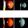
Left: Advanced visually significant posterior subcapsular cataract in the right eye at initial presentation. Right: Advanced visually significant posterior subcapsular cataract in the left eye at initial presentation. (Reproduced with permission from Sharma, P, Vasavada, AR. J Cataract Refract Surg 27:789–794, 2001) Left: Progressing centripetal regression of the posterior subcapsular cataract in the right eye 3 weeks after the initial presentation. Right: Irregular pattern of regression of the posterior subcapsular cataract in the left eye 3 weeks after the initial presentation. (Reproduced with permission from Sharma, P, Vasavada, AR. J Cataract Refract Surg 27:789–794, 2001)
FIGURE 2
FIGURE 2
Nonproliferative diabetic retinopathy with clinically significant macular edema. (Reproduced with permission from The Wills Eye Manual: Office and Emergency Room Diagnosis and Treatment of Eye Disease, 5th edition; Ehlers and Shah, editors, Wolters Kluwer|Lippincott Williams and Wilkins, Philadelphia, 2008)
FIGURE 3
FIGURE 3
Proliferative retinopathy with disc vessels, gliosis, and traction retinal detachment superiorly. (Reproduced with permission from Naji, A. and Vernon, SA. J R Soc Med 96:266–272, 2003)
FIGURE 4
FIGURE 4
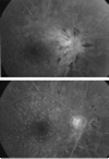
Left eye. (Top) Late-phase fluorescein angiogram shows leakage from the optic nerve head with surrounding peripapillary hemorrhages. (Bottom) Late-phase fluorescein angiogram 2 weeks later shows almost complete resolution of leakage from the optic nerve head with decrease in the hemorrhages. Vision improved from counting fingers to 20/50. (Reproduced with permission from Al-Haddad, CE, Jurdi, FA, and Bashshur, ZF. Am J Ophthalmol 137:1151–1153, 2004)
FIGURE 5
FIGURE 5
Right eye. (Top) Late-phase fluorescein angiogram shows leakage from the optic nerve head with counting fingers vision. (Bottom) Two weeks later, there is significant decrease in the leakage on the late-phase angiogram and improvement in vision to 20/40. (Reproduced with permission from Al-Haddad, CE, Jurdi, FA, and Bashshur, ZF. Am J Ophthalmol 137:1151–1153, 2004)

References

    1. CDC. Diabetes Success and opportunities for population-based prevention and control: at a glance. 2009
    1. Congdon N, Freidman D, Lietman T. Important Causes of Visual Impairment in the World Today. JAMA. 2003;290:2057–2060. - PubMed
    1. Waite JH, Beetham WP. The visual mechanism in diabetes mellitus: A comprehensive study of 2002 diabetics and 457 non- diabetics for control. New Engl. J. Med. 1935;212:367–379. 429–443.
    1. Rocha G, Garza G, Font RL. Orbital pathology associated with diabetes mellitus. Int. Ophthalmol. Clin. 1998;38(2):169–179. - PubMed
    1. Herse PR. A review of manifestations of diabetes mellitus in the anterior eye and cornea. Am. J. Optom. Physiol. Optics. 1988;65(3):224–230. - PubMed

WEBSITES

    1. (70) CDC. National Diabetes Fact Sheet. 2007 http://www.cdc.gov/diabetes/pubs/estimates07.htm.

LinkOut - more resources