

Collagen and elastic fibers in odontogenic entities: analysis using light and confocal laser microscopic methods
- PMID: 21760864
- PMCID: PMC3134956
- DOI: 10.2174/1874210601105010116
Collagen and elastic fibers in odontogenic entities: analysis using light and confocal laser microscopic methods
Abstract
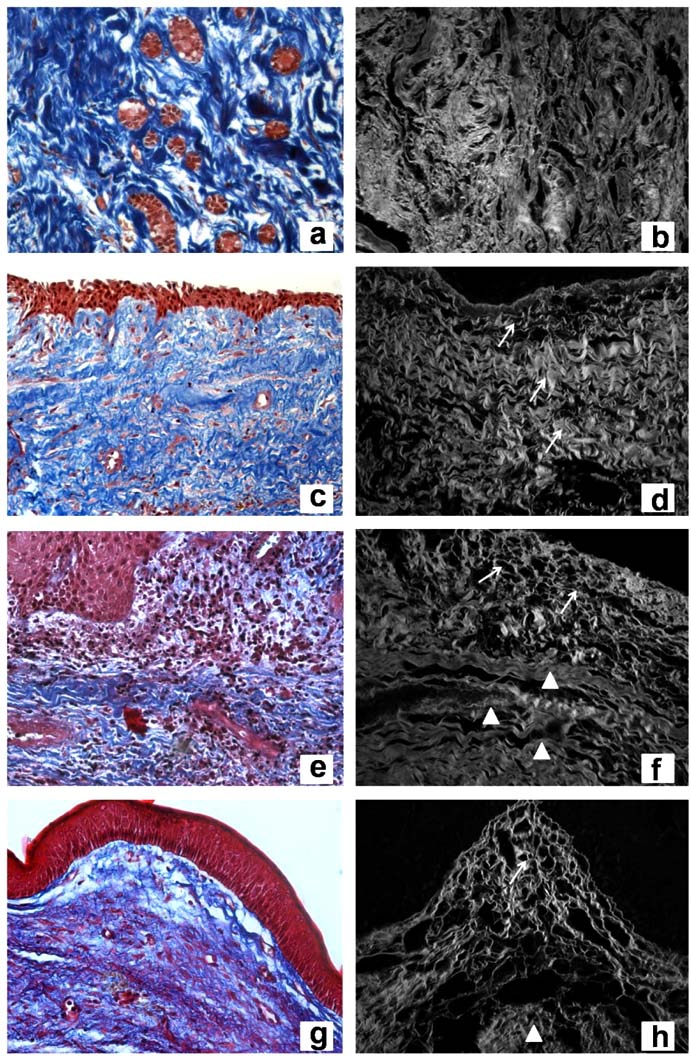
Dentigerous cyst (DC) and keratocystic odontogenic tumor (KOT) are odontogenic lesions arising from epithelial elements, such as those observed in dental follicles (DF), that have been part of the tooth forming apparatus. These lesions show different clinical and histological characteristics, as well as distinct biological behavior. This study aimed to qualify and quantify collagen and elastic fibers by means of histochemical techniques with light and confocal laser microscopic methods in three odontogenic entities. Eleven DF, 13 DC (n=10 with inflammation, n=3 without inflammation) and 13 KOT were processed to the following techniques: Hematoxylin and Eosin, Masson's Trichrome, Picrosirius, Direct Blue, and Orcein. DF and DC without inflammation exhibited collagen with similar characteristics: no parallel pattern of fiber orientation, thick fibers with dense arrangement, and absence of distinct layers. A comparison between DC with inflammation and KOT revealed similar collagen organization, showing distinct layers: thin collagen fibers with loose arrangement near the epithelium and thick fibers with dense arrangement in distant areas. The only difference found was that KOT exhibited a parallel collagen orientation in relation to the odontogenic epithelia. It may be suggested that the connective tissue of DC is a reactive tissue, inducing an expansive growth associated with fluid accumulation and inflammatory process, which in turn may be present as part of the lesion itself. In KOT, loosely arranged collagen may be associated with the behavior of the neoplastic epithelium.
Keywords: Dental follicle; confocal laser microscopy.; dentigerous cyst; extracellular matrix; keratocystic odontogenic tumor.
Figures
References
-
- Kim J, Ellis G. Dental follicular tissue: misinterpretation as odontogenic tumors. J Oral Maxillofac Surg. 1993;51:762–7. - PubMed
-
- Rakprasitkul S. Pathologic changes in the pericoronal tissues of unerupted third molars. Quintessence Int. 2001;32:633–8. - PubMed
-
- Curran A, Damm D, Drummond J. Pathologically significant pericoronal lesions in adults: histopathologic evaluation. J Oral Maxillofac Surg. 2002;60:613–7. - PubMed
-
- Saraçoglu U, Kurt B, Günhan , Güven O. MIB-1 expression in odontogenic epithelial rests, epithelium of health oral mucosa and epithelium of selected odontogenic cysts: an immunohistochemical study. Int. J Oral Maxillofac Surg. 2005;34:432–43. - PubMed
-
- Ide F, Obara K, Yamada H, et al. Hamartomatous proliferations of odontogenic epithelium within the jaws: a potential histogenetic source of intraosseous epithelial odontogenic tumors. J Oral Pathol Med. 2007;36:229–35. - PubMed
LinkOut - more resources
Full Text Sources
Miscellaneous