N-terminal prolactin-derived fragments, vasoinhibins, are proapoptoptic and antiproliferative in the anterior pituitary
- PMID: 21760910
- PMCID: PMC3131298
- DOI: 10.1371/journal.pone.0021806
N-terminal prolactin-derived fragments, vasoinhibins, are proapoptoptic and antiproliferative in the anterior pituitary
Abstract
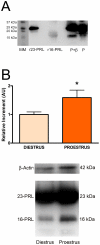
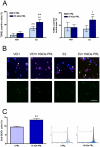
The anterior pituitary is under a constant cell turnover modulated by gonadal steroids. In the rat, an increase in the rate of apoptosis occurs at proestrus whereas a peak of proliferation takes place at estrus. At proestrus, concomitant with the maximum rate of apoptosis, a peak in circulating levels of prolactin is observed. Prolactin can be cleaved to different N-terminal fragments, vasoinhibins, which are proapoptotic and antiproliferative factors for endothelial cells. It was reported that a 16 kDa vasoinhibin is produced in the rat anterior pituitary by cathepsin D. In the present study we investigated the anterior pituitary production of N-terminal prolactin-derived fragments along the estrous cycle and the involvement of estrogens in this process. In addition, we studied the effects of a recombinant vasoinhibin, 16 kDa prolactin, on anterior pituitary apoptosis and proliferation. We observed by Western Blot that N-terminal prolactin-derived fragments production in the anterior pituitary was higher at proestrus with respect to diestrus and that the content and release of these prolactin forms from anterior pituitary cells in culture were increased by estradiol. A recombinant preparation of 16 kDa prolactin induced apoptosis (determined by TUNEL assay and flow cytometry) of cultured anterior pituitary cells and lactotropes from ovariectomized rats only in the presence of estradiol, as previously reported for other proapoptotic factors in the anterior pituitary. In addition, 16 kDa prolactin decreased forskolin-induced proliferation (evaluated by BrdU incorporation) of rat total anterior pituitary cells and lactotropes in culture and decreased the proportion of cells in S-phase of the cell cycle (determined by flow cytometry). In conclusion, our study indicates that the anterior pituitary production of 16 kDa prolactin is variable along the estrous cycle and increased by estrogens. The antiproliferative and estradiol-dependent proapoptotic actions of this vasoinhibin may be involved in the control of anterior pituitary cell renewal.
Conflict of interest statement
Figures


Similar articles
-
Estrogens up-regulate the Fas/FasL apoptotic pathway in lactotropes.Endocrinology. 2005 Nov;146(11):4737-44. doi: 10.1210/en.2005-0279. Epub 2005 Aug 11. Endocrinology. 2005. PMID: 16099864 Free PMC article.
-
Estrogens sensitize anterior pituitary gland to apoptosis.Am J Physiol Endocrinol Metab. 2004 Oct;287(4):E767-71. doi: 10.1152/ajpendo.00052.2004. Epub 2004 Jun 1. Am J Physiol Endocrinol Metab. 2004. PMID: 15172886
-
TNF-alpha induces apoptosis of lactotropes from female rats.Endocrinology. 2002 Sep;143(9):3611-7. doi: 10.1210/en.2002-220377. Endocrinology. 2002. PMID: 12193577
-
Role of estrogens in anterior pituitary gland remodeling during the estrous cycle.Front Horm Res. 2010;38:25-31. doi: 10.1159/000318491. Epub 2010 Jul 5. Front Horm Res. 2010. PMID: 20616492 Review.
-
Anterior pituitary cell renewal during the estrous cycle.Front Horm Res. 2006;35:9-21. doi: 10.1159/000094260. Front Horm Res. 2006. PMID: 16809919 Review.
Cited by
-
Principles of the prolactin/vasoinhibin axis.Am J Physiol Regul Integr Comp Physiol. 2015 Nov 15;309(10):R1193-203. doi: 10.1152/ajpregu.00256.2015. Epub 2015 Aug 26. Am J Physiol Regul Integr Comp Physiol. 2015. PMID: 26310939 Free PMC article. Review.
-
Prolactin induces apoptosis of lactotropes in female rodents.PLoS One. 2014 May 23;9(5):e97383. doi: 10.1371/journal.pone.0097383. eCollection 2014. PLoS One. 2014. PMID: 24859278 Free PMC article.
-
Is prolactin receptor signaling a target in dopamine-resistant prolactinomas?Front Endocrinol (Lausanne). 2023 Jan 12;13:1057749. doi: 10.3389/fendo.2022.1057749. eCollection 2022. Front Endocrinol (Lausanne). 2023. PMID: 36714572 Free PMC article. Review.
References
-
- Yin P, Arita J. Proestrus surge of Gonadotropin-releasing hormone secretion inhibits apoptosis of anterior pituitary cells in cycling female rats. Neuroendocrinology. 2002;76:272–282. - PubMed
-
- Hashi A, Mazawa S, Chen S, Kato J, Arita J. Pentobarbital anesthesia during the proestrous afternoon blocks lactotroph proliferation occurring on estrus in female rats. Endocrinology. 1995;136:4665–4671. - PubMed
-
- Candolfi M, Zaldivar V, De Laurentiis A, Jaita G, Pisera D, et al. TNF-alpha induces apoptosis of lactotropes from female rats. Endocrinology. 2002;143:3611–3617. - PubMed
-
- Radl DB, Zárate S, Jaita G, Ferraris J, Zaldivar V, et al. Apoptosis of lactotrophs induced by D2 receptor activation is estrogen dependent. Neuroendocrinology. 2008;88:43–52. - PubMed
Publication types
MeSH terms
Substances
LinkOut - more resources
Full Text Sources

