

The default mode network in healthy aging and Alzheimer's disease
- PMID: 21760988
- PMCID: PMC3132539
- DOI: 10.4061/2011/535816
The default mode network in healthy aging and Alzheimer's disease
Abstract
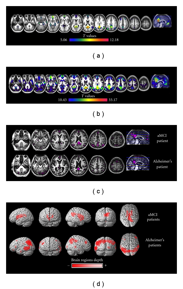
In the past decade, a "default mode network" (DMN) has been highlighted in neuroimaging studies as a set of brain regions showing increased activity in task-free state compared to cognitively demanding task, and synchronized activity at rest. Changes within this network have been described in healthy aging as well as in Alzheimer's disease (AD) and populations at risk for AD, that is, amnestic Mild Cognitive Impairment (aMCI) patients and APOE-ε4 carriers. This is of particular interest in the context of early diagnosis and more generally for our understanding of the physiopathological mechanisms of AD. This paper gives an overview of the anatomical and physiological characteristics of this network as well as its relationships with cognition, before focusing on changes in the DMN over normal aging and Alzheimer's disease. While perturbations of the DMN have been consistently reported, especially within the posterior cingulate, further studies are needed to understand their clinical implication.
Figures
References
-
- Ghatan PH, Hsieh JC, Wirsen-Meurling A, et al. Brain activation induced by the perceptual maze test: a PET study of cognitive performance. NeuroImage. 1995;2(2):112–124. - PubMed
-
- Hutchinson M, Schiffer W, Joseffer S, et al. Task-specific deactivation patterns in functional magnetic resonance imaging. Magnetic Resonance Imaging. 1999;17(10):1427–1436. - PubMed
-
- Gusnard DA, Raichle ME. Searching for a baseline: functional imaging and the resting human brain. Nature Reviews Neuroscience. 2001;2(10):685–694. - PubMed
-
- McKiernan KA, Kaufman JN, Kucera-Thompson J, Binder JR. A parametric manipulation of factors affecting task-induced deactivation in functional neuroimaging. Journal of Cognitive Neuroscience. 2003;15(3):394–408. - PubMed
LinkOut - more resources
Full Text Sources
Other Literature Sources
Miscellaneous
