. 2011 Aug;2(4):239-48.
doi: 10.1007/s12672-011-0074-6.

Alcohol exposure in utero leads to enhanced prepubertal mammary development and alterations in mammary IGF and estradiol systems

Affiliations

Alcohol exposure in utero leads to enhanced prepubertal mammary development and alterations in mammary IGF and estradiol systems

Tiffany A Polanco et al. Horm Cancer. 2011 Aug.

Abstract

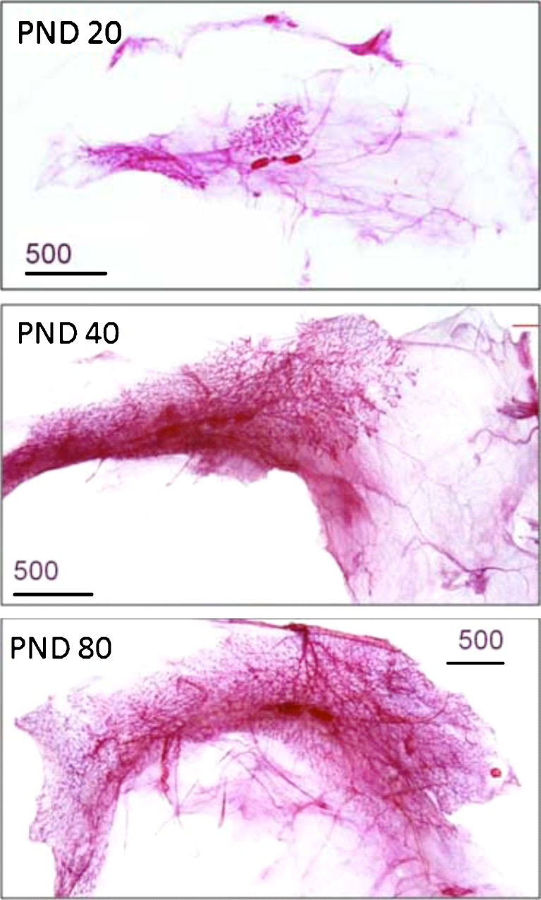
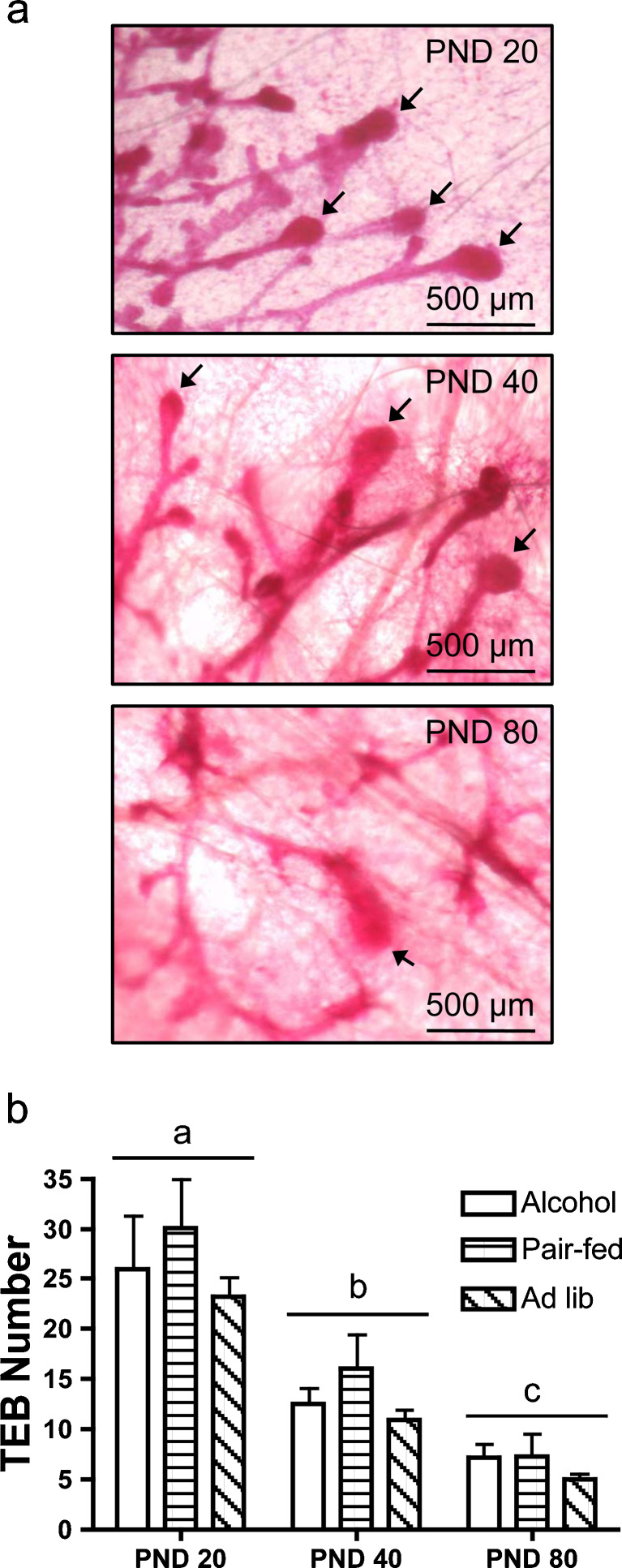
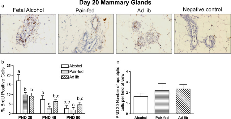
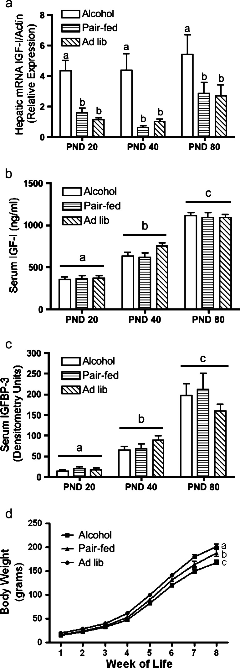
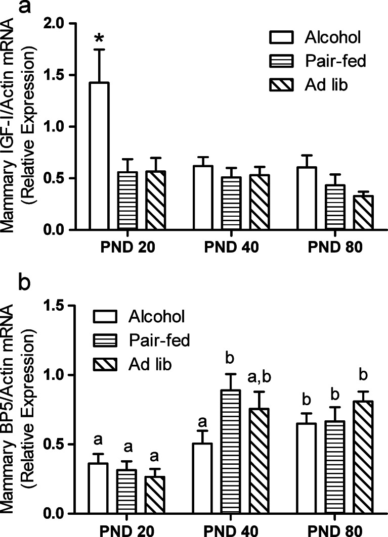
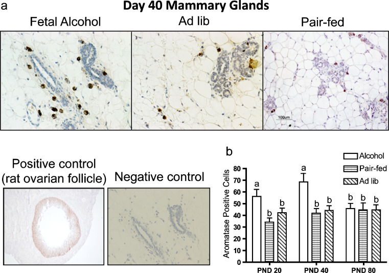
Exposure to alcohol during fetal development increases susceptibility to mammary cancer in adult rats. This study determined if early changes in mammary morphology and the insulin-like growth factor (IGF)/estradiol axis are involved in the mechanisms that underlie this increased susceptibility. Pregnant Sprague-Dawley rats were fed a liquid diet containing 6.7% ethanol (alcohol), an isocaloric liquid diet (pair-fed), or rat chow ad libitum from days 11 to 21 of gestation. At birth, female pups were cross-fostered to ad libitum-fed control dams. Offspring were euthanized at postnatal days (PND) 20, 40, or 80. Animals were injected with BrdU before euthanasia, then mammary glands, serum, and livers were collected. Mammary glands from animals exposed to alcohol in utero displayed increased epithelial cell proliferation and aromatase expression at PND 20 and 40. Mammary IGF-I mRNA was higher in alcohol-exposed animals relative to controls at PND 20, while mammary IGFBP-5 mRNA was lower in this group at PND 40. Hepatic IGF-I mRNA expression was increased at all time points in alcohol-exposed animals, however, circulating IGF-I levels were not altered. These data indicate that alcohol exposure in utero may advance mammary development via the IGF and estradiol systems, which could contribute to increased susceptibility to mammary cancer later in life.


Conflict of interest statement

The authors declare that they have no conflict of interest.

Figures

Fig. 1
Fig. 1
Gross mammary gland morphology is not affected by alcohol exposure in utero. Pups exposed to alcohol in utero and controls not exposed were euthanized at PND 20, 40, or 80. The fifth inguinal mammary gland was processed for whole-mount analysis. Mammary density and length of the ductal structure were not affected by treatment at any time point; therefore, representative whole mounts are shown without respect to treatment
Fig. 2
Fig. 2
Terminal end bud (TEB) numbers decrease with time, but are not affected by in utero alcohol exposure. Pups exposed to alcohol in utero and controls not exposed were euthanized at PND 20, 40, or 80. The fifth inguinal mammary gland was processed for whole-mount analysis, and total TEBs were counted for each gland. a Digital images of whole mounts taken at ×8 magnification. Arrows represent TEBs. b Bars represent mean ± SEM of total TEBs per gland
Fig. 3
Fig. 3
Epithelial cell proliferation is higher in animals exposed to alcohol in utero at PND 20 and 40. Mammary gland sections were prepared and stained for BrdU by immunohistochemistry as described in Materials and Methods. a Representative sections stained for BrdU. The negative control represents a section stained with an isotype IgG antibody. b BrdU incorporation was quantified by counting BrdU-stained cells and expressing them as a percent of total epithelial cells. For each mammary gland, two serial sections were counted and averaged. Total epithelial cells in each section were counted for PND 20, while up to 1,000 cells per section were counted for PND 40 and 80. Bars represent mean ± SEM with different letters denoting a significant difference within each treatment group. A two-way ANOVA was performed with a Bonferroni post hoc test at the level of α = 0.05 (n = 10, 9, and 13 for alcohol, pair-fed, and ad lib groups, respectively, at each time point). d Number of apoptotic cells per field of view
Fig. 4
Fig. 4
Hepatic IGF-I mRNA is increased in animals exposed to alcohol in utero, while serum IGF-I and IGFBP-3 increase over time in all treatment groups. Hepatic tissue and serum was collected from animals exposed to alcohol and controls not exposed and prepared as described in Materials and Methods. a Hepatic IGF-I mRNA expression was assayed by quantitative RT-PCR (P < 0.05). b Circulating IGF-I levels were assayed by ELISA (P < 0.001). c Serum IGFBP-3 levels were assayed by ligand blot (P < 0.0001). d Weekly body weight recordings (P < 0.0001). Bars represent mean ± SEM with different letters denoting significant differences. A two-way ANOVA was performed in ac with a Bonferroni post-test at the level of α = 0.05 (n = 10, 9, or 13 for alcohol, pair-fed, and ad lib groups, respectively, at each time point). A repeated-measures one-way ANOVA was performed in d with a Newman–Keuls post-test at the level of α = 0.05 (during weeks 1–4, n = 30, 28, and 40; during weeks 5 and 6, n = 20, 18, and 27; and during weeks 7 and 8, n = 10, 9, and 13 for alcohol, pair-fed, and ad lib groups, respectively)
Fig. 5
Fig. 5
IGF-I mRNA is significantly higher at PND 20 and IGFBP-5 mRNA increased later in the mammary glands of alcohol-exposed animals. Mammary gland tissue was collected; RNA was isolated, and quantitative PCR was performed as described in Materials and Methods. a Mammary gland IGF-I mRNA expression with asterisk denoting significant difference (P < 0.001). b Mammary gland IGFBP-5 mRNA expression with different letters denoting significant differences (P < 0.05). Bars represent mean ± SEM. A two-way ANOVA was performed with a Bonferroni post-test at the level of α = 0.05 (n = 10, 9, or 13 for alcohol, pair-fed, and ad lib groups, respectively, at each time point)
Fig. 6
Fig. 6
Aromatase expression is increased in the mammary glands of animals exposed to alcohol at PND 20 and 40. Mammary glands were prepared and stained for aromatase as described in Materials and Methods. a Representative sections of aromatase-stained cells. Negative control represents a section stained with an isotype IgG antibody. b Aromatase-positive cells were quantified by counting three fields of view and totaling the numbers for each section. Bars represent mean ± SEM of aromatase-positive cells per gland with different letters denoting significant differences within a treatment group (P < 0.05). A two-way ANOVA was performed with a Bonferroni post hoc test at the level of α = 0.05 (n = 10, 9, or 13 for alcohol, pair-fed, and ad lib groups respectively, at each time point)

Similar articles

Cited by

References

    1. Centers for Disease Control (2004) Alcohol consumption among women who are pregnant or who might become pregnant-United States. In Morbidity and Mortality Weekly Report 53 (50):1178–1181 - PubMed
    1. Sampson PD, Streissguth AP, Bookstein FL, Little RE, Clarren SK, Dehaene P, Hanson JW, Graham JM., Jr Incidence of fetal alcohol syndrome and prevalence of alcohol-related neurodevelopmental disorder. Teratology. 1997;56(5):317–326. doi: 10.1002/(SICI)1096-9926(199711)56:5<317::AID-TERA5>3.0.CO;2-U. - DOI - PubMed
    1. Barker DJ. In utero programming of chronic disease. Clin Sci (Lond) 1998;95(2):115–128. doi: 10.1042/CS19980019. - DOI - PubMed
    1. Barker DJ. The origins of the developmental origins theory. J Intern Med. 2007;261(5):412–417. doi: 10.1111/j.1365-2796.2007.01809.x. - DOI - PubMed
    1. Hilakivi-Clarke L, Cabanes A, de Assis S, Wang M, Khan G, Shoemaker WJ, Stevens RG. In utero alcohol exposure increases mammary tumorigenesis in rats. Br J Cancer. 2004;90(11):2225–2231. - PMC - PubMed

Publication types

MeSH terms

LinkOut - more resources