. 2011 Nov;16(6):607-19.
doi: 10.1007/s12192-011-0270-x. Epub 2011 Jul 15.

Endoplasmic reticulum stress or mutation of an EF-hand Ca(2+)-binding domain directs the FKBP65 rotamase to an ERAD-based proteolysis

Affiliations

Endoplasmic reticulum stress or mutation of an EF-hand Ca(2+)-binding domain directs the FKBP65 rotamase to an ERAD-based proteolysis

Lindsey A Murphy et al. Cell Stress Chaperones. 2011 Nov.

Abstract

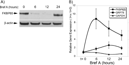
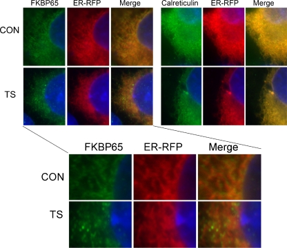
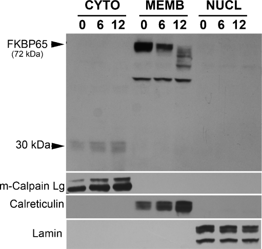
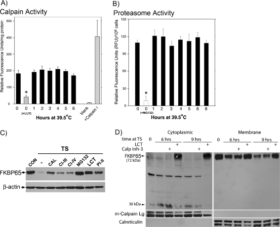
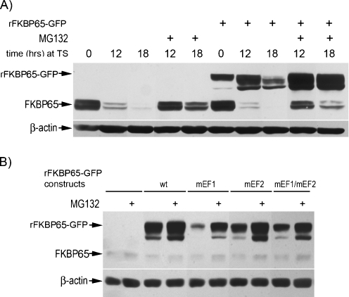
FKBP65 is an endoplasmic reticulum (ER)-localized chaperone and rotamase, with cargo proteins that include tropoelastin and collagen. In humans, mutations in FKBP65 have recently been shown to cause a form of osteogenesis imperfecta (OI), a brittle bone disease resulting from deficient secretion of mature type I collagen. In this work, we describe the rapid proteolysis of FKBP65 in response to ER stress signals that activate the release of ER Ca(2+) stores. A large-scale screen for stress-induced cellular changes revealed FKBP65 proteins to decrease within 6-12 h of stress activation. Inhibiting IP(3)R-mediated ER Ca(2+) release blocked this response. No other ER-localized chaperone and folding mediators assessed in the study displayed this phenomenon, indicating that this rapid proteolysis of folding mediator is distinctive. Imaging and cellular fractionation confirmed the localization of FKBP65 (72 kDa glycoprotein) to the ER of untreated cells, a rapid decrease in protein levels following ER stress, and the corresponding appearance of a 30-kDa fragment in the cytosol. Inhibition of the proteasome during ER stress revealed an accumulation of FKBP65 in the cytosol, consistent with retrotranslocation and a proteasome-based proteolysis. To assess the role of Ca(2+)-binding EF-hand domains in FKBP65 stability, a recombinant FKBP65-GFP construct was engineered to ablate Ca(2+) binding at each of two EF-hand domains. Cells transfected with the wild-type construct displayed ER localization of the FKBP65-GFP protein and a proteasome-dependent proteolysis in response to ER stress. Recombinant FKBP65-GFP carrying a defect in the EF1 Ca(2+)-binding domain displayed diminished protein in the ER when compared to wild-type FKBP65-GFP. Proteasome inhibition restored mutant protein to levels similar to that of the wild-type FKBP65-GFP. A similar mutation in EF2 did not confer FKBP65 proteolysis. This work supports a model in which stress-induced changes in ER Ca(2+) stores induce the rapid proteolysis of FKBP65, a chaperone and folding mediator of collagen and tropoelastin. The destruction of this protein may identify a cellular strategy for replacement of protein folding machinery following ER stress. The implications for stress-induced changes in the handling of aggregate-prone proteins in the ER-Golgi secretory pathway are discussed. This work was supported by grants from the National Institutes of Health (R15GM065139) and the National Science Foundation (DBI-0452587).


Figures

Fig. 1
Fig. 1
Endoplasmic reticulum stress induces FKBP65 proteolysis. a Representative immunoblot results from Powerblot™ analysis of lysates from tsBN7 cells. Cells were exposed to 39.5°C (TS) or tunicamycin (TUN, 1 μM) for 24 h. b Immunoblot analysis of select ER-localized folding mediators following a 6-h exposure to ER stressor; staurosporine (STS, 1 μM), an ER-independent apoptosis activator; or cyclohexmide (CHX, 10 μM). c A time course of FKBP65 proteolysis following activation of ER stress with either TS or TUN treatment. Note the appearance of underglycosylated FKBP65 after 18 h of stress exposure. d Pro-apoptotic ER stress signaling can be inhibited by blocking ER Ca2+ release (2-APB, 50 μM) or cellular calpain proteases (calpeptin, 100 μM). Significant differences between TS-treated cells and TS-treated cells with inhibitors were observed at 36 and 48 h (ANOVA statistical test, p < 0.05). e Inhibition of calpains or ER Ca2+ release is sufficient to partially block FKBP65 proteolysis following ER stress
Fig. 2
Fig. 2
ER stress does not inhibit FKBP65 expression. a In human SH-SY5Y cells, ER stress was induced using Brefeldin A (20 μM), activating a rapid depletion of FKBP65, similar to that seen in fibroblast cell lines. b Quantitative reverse transcription followed by polymerase chain reaction (RT/PCR) was used to examine the effect of ER stress upon expression of FKBP65 and GRP78. The prevalence of each transcript was determined by normalizing PCR amplification signal relative to a known standard for each gene. Normalized to t = 0, ER stress induces an eight-fold increase in GRP78 expression and a moderate increase in FKBP65 expression
Fig. 3
Fig. 3
ER stress diminishes FKBP65 co-localization with the ER. tsBN7 fibroblasts were transfected with pER-RFP (Invitrogen) to label the endoplasmic reticulum with RFP. Immunologic detection of FKBP65 or calreticulin involved a primary antibody detected with an Alexa 488-conjugated secondary antibody (Molecular Probes; Eugene, OR, USA). A 12-h TS treatment was used to induce ER stress in these tsBN7 fibroblasts. FKBP65, but not calreticulin, displayed diminished signal intensity and ER localization following TS treatment
Fig. 4
Fig. 4
ER stress induces a decrease of FKBP65 localization to membrane fractions and evidence of a cytosolic cleavage product. Subcellular fractionation was performed using cell lysates from tsBN7 cells exposed to TS for 0, 6, or 12 h. Fractions generated were cytosolic, membrane (ER, mitochondria), and nuclear fractions. The integrity of each fraction was determined by examining markers for each cellular compartment (cytosol, calpain; ER, calreticulin; nucleus, lamin). Following ER stress, FKBP65 within the ER diminishes in intensity, while a 30-kDa FKBP65 antibody-reactive protein increases in intensity
Fig. 5
Fig. 5
Proteasome inhibition blocks FKBP65 proteolysis in the cytosol. a, b An assay of cellular calpain and proteasome activities following ER stress activation in tsBN7 fibroblasts. In each case, the cleavage of fluorogenic substrates for protease activity was quantitatively measured using 96-well plate assays. Both activities are present in these cells, but ER stress does not significantly alter activity at acute time points. Statistical significance was determined using ANOVA with a Tukey's all pairs post hoc test (p < 0.05). c Assessment of ER stress-induced proteolysis of FKBP65 in the presence of different membrane permeable calpain or proteasome inhibitors (6-h time point). Proteasome inhibitors (MG132, 5 μM; lactacystin (LCT), 5 μM; Proteasome Inhib. II, 5 μM) were highly effective at inhibiting FKBP65 proteolysis, while calpain inhibitors partially inhibited this phenomenon (calpeptin (CAL), 100 μM; Calpain Inhib. III/IV, 10 μM). d Subcellular fractionation was performed using cell lysates from tsBN7 cells exposed to TS for 6 or 9 h in the presence or absence of calpain inhibitor (CI-III, 10 μM) or proteasome inhibitor (LCT, 5 μM). The quality of cellular fractions was assessed using m-calpain as a marker of the cytosol and calreticulin as an ER-specific marker for the membrane fraction. To enable visualization of the 30-kDa band, the cytosolic FKBP65 panel was exposed 5× longer than the membrane panel in 5D. All other paired panels were exposed to film for identical time periods. TS treatment generated an accumulation of cytosolic FBKP10 when the proteasome was inhibited, but not when calpain was inhibited
Fig. 6
Fig. 6
The EF-hand Ca2+-binding domain regulates FKBP65 stability in the ER. a EF-hand Ca2+-binding domains from several proteins, each displaying acidic aspartate (d) and glutamic acid (e) residues at each end of the domain to facilitate Ca2+ binding at the intersection of two α-helical domains. b An FKBP65-GFP fusion protein was created with an ER retention signal (HEEL), a flexible linker (GSGS), and EE → KK mutations in each EF-hand domain (EF1 and EF2). c Expression of the non-mutant rFKBP65-GFP construct in cultured tsBN7 cells reveals co-localization with ER-RFP consistent with localization to the ER. d Expression of non-mutant rFKBP65-GFP, mEF1, mEF2, and mEF1/mEF2 constructs in tsBN7 fibroblasts. Fractionation of transfected cells reveals the recombinant proteins to display localization within the membrane fraction, consistent with ER localization. Mutation of the EF1 Ca2+-binding domain results in diminished rFKBP65-GFP protein within the cell despite cells being transfected with identical amounts of plasmid DNA and equal loading of protein lysates
Fig. 7
Fig. 7
Proteolysis of EF-hand mutant FKBP65 is mediated by the proteasome. a Cultured tsBN7 cells expressing rFKBP65-GFP display an ER stress-induced proteolysis of the recombinant protein, similar to the native FKBP65 protein. Inhibition of the proteasome inhibited this proteolysis (MG132, 5 μM). b Proteasome inhibition (12-h exposure) is sufficient to restore normal cellular accumulation of rFKBP65-GFP proteins carrying mutations in the EF-hand Ca2+-binding domains. To generate the final figures (a, b), some lanes were reordered, though all bands shown in each panel are from a photograph of a single auto-radiographic film

Similar articles

Cited by

References

    1. Alanay Y, et al. Mutations in the gene encoding the RER protein FKBP65 cause autosomal-recessive osteogenesis imperfecta. Am J Hum Genet. 2010;86:551–559. doi: 10.1016/j.ajhg.2010.02.022. - DOI - PMC - PubMed
    1. Bandopadhyay R, Belleroche J. Pathogenesis of Parkinson's disease: emerging role of molecular chaperones. Trends Mol Med. 2010;16:27–36. doi: 10.1016/j.molmed.2009.11.004. - DOI - PubMed
    1. Brewster JL, Linseman DA, Bouchard RJ, Loucks FA, Precht TA, Esch EA, Heidenreich KA. Endoplasmic reticulum stress and trophic factor withdrawal activate distinct signaling cascades that induce glycogen synthase kinase 3beta and a caspase-9-dependent apoptosis in cerebellar granule neurons. Mol Cell Neurosci. 2006;32:242–253. doi: 10.1016/j.mcn.2006.04.006. - DOI - PubMed
    1. Butterfield DA, Abdul HM, Opil W, Newman SF, Joshi G, Ansari MA, Sultana R. Pin1 in Alzheimer's disease. J Neurochem. 2006;98:1697–1706. doi: 10.1111/j.1471-4159.2006.03995.x. - DOI - PubMed
    1. Cheung KL, Bates M, Ananthanarayanan VS. Effect of FKBP65, a putative elastin chaperone, on the coacervation of tropoelastin in vitro. Biochem Cell Biol. 2010;88:917–925. doi: 10.1139/O10-137. - DOI - PubMed

Publication types