. 2011 Oct;356(1-2):209-16.
doi: 10.1007/s11010-011-0961-8. Epub 2011 Jul 15.

CK2α is essential for embryonic morphogenesis

Affiliations

CK2α is essential for embryonic morphogenesis

Isabel Dominguez et al. Mol Cell Biochem. 2011 Oct.

Abstract

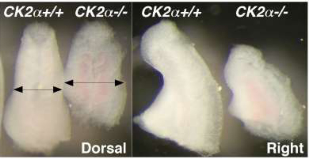
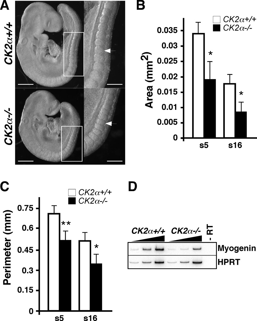
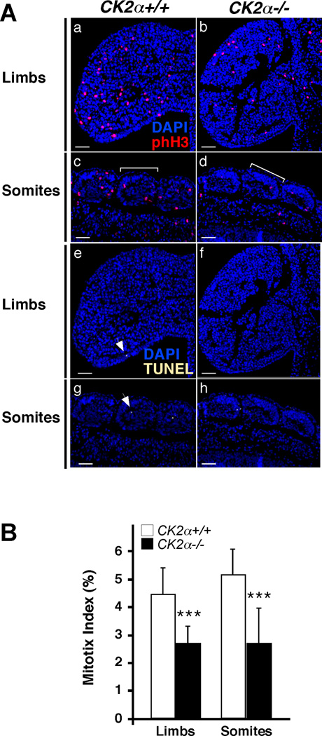
CK2 is a highly conserved serine-threonine kinase involved in biological processes such as embryonic development, circadian rhythms, inflammation, and cancer. Biochemical experiments have implicated CK2 in the control of several cellular processes and in the regulation of signal transduction pathways. Our laboratory is interested in characterizing the cellular, signaling, and molecular mechanisms regulated by CK2 during early embryonic development. For this purpose, animal models, including mice deficient in CK2 genes, are indispensable tools. Using CK2α gene-deficient mice, we have recently shown that CK2α is a critical regulator of mid-gestational morphogenetic processes, as CK2α deficiency results in defects in heart, brain, pharyngeal arch, tail bud, limb bud, and somite formation. Morphogenetic processes depend upon the precise coordination of essential cellular processes in which CK2 has been implicated, such as proliferation and survival. Here, we summarize the overall phenotype found in CK2α (-/- ) mice and describe our initial analysis aimed to identify the cellular processes affected in CK2α mutants.


Figures

Fig. 1
Fig. 1. Photographs of tail buds of E9.5 WT and CK2α−/− embryos
Dorsal and right views of CK2α+/+ and CK2α−/− embryos at somite pairs (22–23). Arrows show the in increased tail bud width in CK2α−/− embryos. Representative photographs of over 70 embryo litters.
Fig. 2
Fig. 2. Phenotype of E9.5 CK2α−/− embryos
(A) Right: right view of 22 somite pair CK2α+/+ and CK2α−/− embryos (Scale bar: 0.5 mm); left: magnified images of the squares on the right (Scale bar: 0.25 mm). Arrow indicates somite number 16. Somites 5 (s5) and 16 (s16) were analyzed for area and perimeter. Histogram showing average somite area (B) and perimeter (C) +/− standard deviation. (*) denotes p<0.05; (**) denotes p<0.005. (D) Decrease in Myogenin transcript expression in CK2α mutant compared to WT embryos by radioactive RT-PCR. Increasing PCR cycles for both CK2α−/− and CK2α+/+ are shown. (-RT) Control minus reverse transcriptase.
Fig. 3
Fig. 3. Proliferation and apoptosis in E9.5 and E10.5 CK2α−/− embryos
(A, B) E9.5 embryos (22–24 somite pairs) were stained by WIHC for phH3 (red, A) and TUNEL (green, B). (C) E10.5 embryos (32–34 somite pairs) were stained by WIHC for phH3 (red) and TUNEL (green). Photographs show bright field (BF), and phH3 and TUNEL staining. Fluorescent images were pseudo-colored. Scale bar: 0.5 mm. Arrowheads point to different embryonic tissues. Abbreviations: fl: forelimb bud; h: heart; hl; hindlimb bud; p1: first pharyngeal arch; s: somite.
Fig. 4
Fig. 4. Head, limb buds and tailbud of E10.5 CK2α−/− embryos
Magnified images of E10.5 embryos (32–34 somite pairs) stained with phH3. Abbreviations: AER: apical ectodermal ridge; di: diencephalon; fl: forelimb bud; hl; hindlimb bud; mes: mesencehalon; met: metencephalon; mye: myelencephalon; p1: first pharyngeal arch; p2: second pharyngeal arch; s: somite; tb: tail bud; tel: telencephalon.
Fig. 5
Fig. 5. Proliferation in E10.5 forelimb buds and somites
(A) Composite phH3 (red)/ DAPI (blue) photographs (a to d) and TUNEL (yellow)/ DAPI (blue) photographs (e to h) of limbs and somites. Bracket indicates one somite. Arrows point to TUNEL+ cells. Fluorescent images were pseudo-colored. Scale bar: 50 µm (B) Histogram represents the average mitotic index (total number of phH3+ cells/ number of DAPI+ cells) +/− standard deviation. A total of 15 sections per tissue were counted from two pairs of CK2α+/+ and CK2α−/− embryos. (***) denotes p<0.0005.

References

    1. Xu X, Toselli PA, Russell LD, Seldin DC. Globozoospermia in mice lacking the casein kinase II alpha' catalytic subunit. Nat Genet. 1999;23(1):118–121. - PubMed
    1. Buchou T, Vernet M, Blond O, Jensen HH, Pointu H, Olsen BB, Cochet C, Issinger OG, Boldyreff B. Disruption of the regulatory beta subunit of protein kinase CK2 in mice leads to a cell-autonomous defect and early embryonic lethality. Mol Cell Biol. 2003;23(3):908–915. - PMC - PubMed
    1. Lou DY, Dominguez I, Toselli P, Landesman-Bollag E, O'Brien C, Seldin DC. The alpha catalytic subunit of protein kinase CK2 is required for mouse embryonic development. Mol Cell Biol. 2008;28(1):131–139. - PMC - PubMed
    1. Moreno-Romero J, Espunya MC, Platara M, Arino J, Martinez MC. A role for protein kinase CK2 in plant development: evidence obtained using a dominant-negative mutant. Plant J. 2008;55(1):118–130. - PubMed
    1. Allada R, Meissner RA. Casein kinase 2, circadian clocks, and the flight from mutagenic light. Mol Cell Biochem. 2005;274(1–2):141–149. - PubMed

Publication types

Substances

LinkOut - more resources