

Immunoguided microdissection techniques
- PMID: 21761293
- PMCID: PMC4794997
- DOI: 10.1007/978-1-61779-163-5_4
Immunoguided microdissection techniques
Abstract
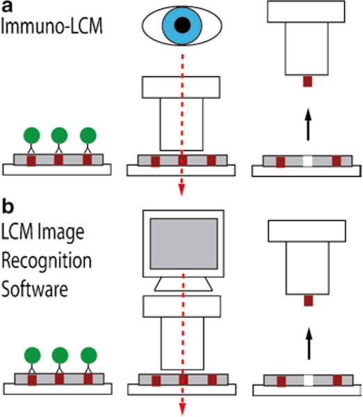
Over the past 15 years, laser-based microdissection has improved the precision by which scientists can procure cells of interest from a heterogeneous tissue section. However, for studies that require a large amount of material (e.g., proteomics) or for cells that are scattered and difficult to identify by standard histological stains, an immunostain-based, automated approach becomes essential. In this chapter, we discuss the use of immunohistochemistry (IHC) and immunofluorescence (IF) to guide the microdissection process via manual and software-driven auto-dissection methods. Although technical challenges still exist with these innovative approaches, we present here methods and protocols to successfully perform immuno-based microdissection on commercially available laser dissection systems.
Figures
References
-
- Coons AH, Creech HJ, Jones RN. Immunological properties of an antibody containing a fluorescent group. Proceedings of the Society for Experimental Biology and Medicine. 1941;47:200–202.
-
- Emmert-Buck MR, Bonner RF, Smith PD, Chuaqui RF, Zhuang Z, Goldstein SR, Weiss RA, Liotta LA. Laser capture microdissection. Science. 1996;274:998–1001. - PubMed
-
- Bonner RF, Emmert-Buck M, Cole K, Pohida T, Chuaqui R, Goldstein S, Liotta LA. Laser capture microdissection: molecular analysis of tissue. Science. 1997;278(1481):1483. - PubMed
-
- Fend F, Kremer M, Quintanilla-Martinez L. Laser capture microdissection: methodical aspects and applications with emphasis on immuno-laser capture microdissection. Pathobiology. 2000;68:209–214. - PubMed
MeSH terms
Substances
Grants and funding
LinkOut - more resources
Full Text Sources
Other Literature Sources
Miscellaneous
