. 2011 Apr;2(2):132-43.
doi: 10.1007/s12672-011-0067-5.

Expression and relevance of TRPS-1: a new GATA transcription factor in breast cancer

Affiliations

Expression and relevance of TRPS-1: a new GATA transcription factor in breast cancer

Jie Qing Chen et al. Horm Cancer. 2011 Apr.

Abstract

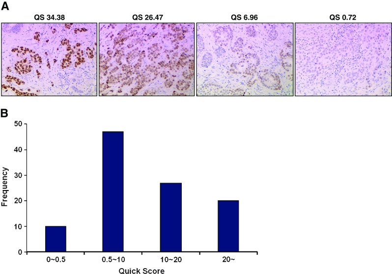
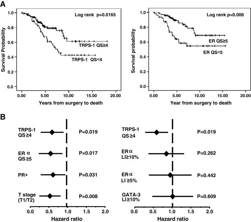
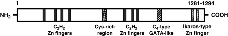
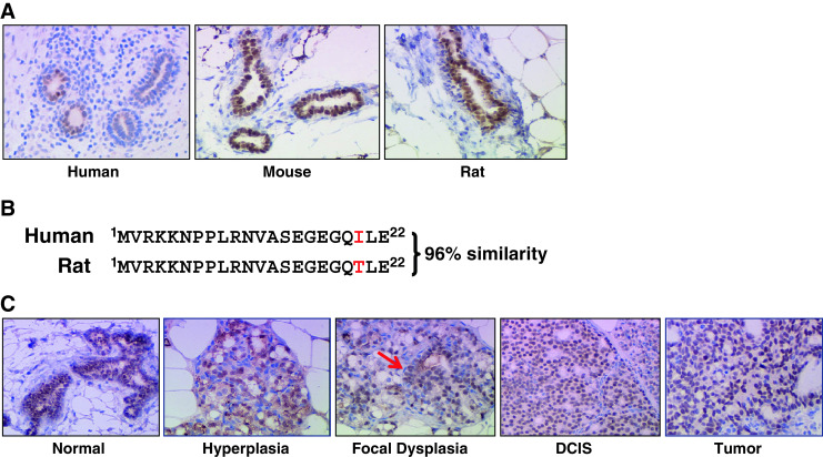
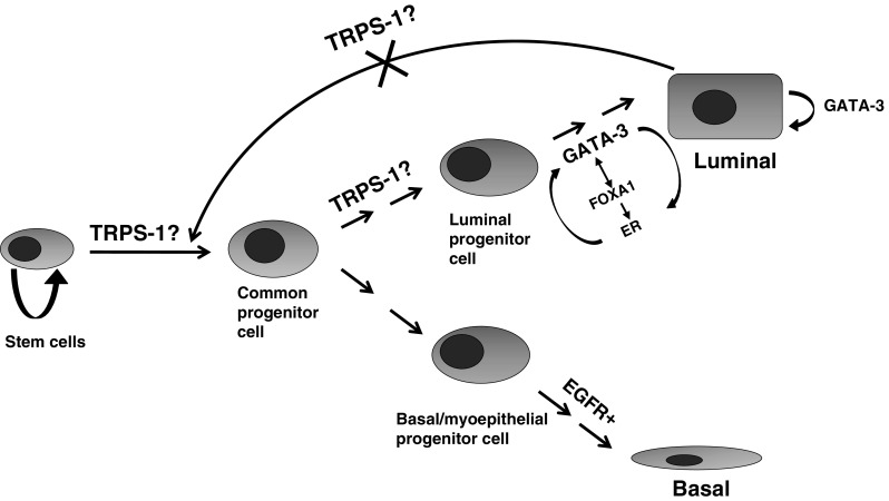
GATA transcription factor family members have been found to play a critical role in the differentiation of many tissue types. For example, GATA-3 has been found to be highly correlated with estrogen receptor α (ER) expression and is emerging as one of the "master regulators" in breast ductal epithelial cell differentiation. Recently, we discovered another GATA family member highly prevalent in breast cancer called the trichorhinophalangeal syndrome-1 gene (TRPS-1). Using a quantitative immunohistochemistry (qIHC) approach, we found that TRPS-1 was significantly correlated with ER, PR, GATA-3, as well as HER2 expression. However, TRPS-1 was also found to be expressed in a high proportion of ER(-) ductal epithelial breast cancers (BCs), indicating that it may act as a ductal epithelial cell-specific transcription factor regulating cell fate at some point in the epithelial cell differentiation pathway. In keeping with this hypothesis, we found that TRPS-1 protein expression in BC above a certain threshold using qIHC correlated with markedly improved overall survival. Cox proportional hazards analysis found that both TRPS-1 and ER expression above critical threshold equally predicted for improved survival. Thus, TRPS-1 may be a powerful new positive prognostic marker in BC, and further IHC studies, as well as examination of its molecular function in ductal epithelial cell differentiation in the breast, are warranted. In this regard, data on the role of TRPS-1 in the differentiation of cells from mesenchymal precursors in other tissues, such as kidney metanephric mesenchymal cells, columnar chondrocytes, and osteoblasts, in mouse models may be useful. Indeed, these studies have found that TRPS-1 is a critical regulator of mesenchymal-to-epithelial cell transition. In the mammary gland, the restricted expression of TRPS-1 in human, mouse, and rat ductal epithelial cells suggests that it may also play a similar role during ductal luminal progenitor/stem cell differentiation. We present a model of TRPS-1 action in which it may act upstream of GATA-3 and ER on an earlier ductal epithelial progenitor cell or mammary stem cell during mammary gland development and also helps prevent reversion of ER(+) BC cells back into mesenchymal-like cells. This model predicts that BCs with low or no TRPS-1 expression may inherently be much less differentiated and more aggressive tumors with less favorable prognosis.


Figures

Fig. 1
Fig. 1
TRPS-1 protein is found at different levels in the nuclei of ductal epithelial cells of BC from different patients using qIHC. a Staining and quantification of TRPS-1 expression using the QS parameter in representative patient BC samples illustrating the heterogeneity of TRPS-1 levels in BC. Magnification is at ×100. b Histogram showing the wide distribution of TRPS-1 QS across the stage II/III BC sample set studied from low to high QS
Fig. 2
Fig. 2
Quantitative IHC reveals that TRPS-1 may be a marker for improved survival in BC. a Kaplan–Meier survival analysis comparing the potential value of TRPS-1 as a marker for improved prognosis to that ER in a cohort of stage II/III BC patients (n = 152). Patients with TRPS-1 QS ≥ 4 had a significantly improved survival versus patients with TRPS-1 QS < 4 (left graph). ER expression was similarly associated with improved outcome with ER QS ≥ 5 group having a significantly longer overall survival than the QS < 5 group (right graph). The p values (p = 0.0165 for TRPS-1; p = 0.006 for ER), calculated using log rank analysis, are shown in each case. b Cox proportional hazards analysis shows TRPS-1 QS ≥ 4 to be significantly associated with better survival with a HR of 0.5 (p = 0.019), similar to ER QS ≥ 5 (HR = 0.48, p = 0.017). Notably, when the labeling index (LI) or percentage positive tumor cells in the samples were scored, ER and GATA-3 LI (≥5% or ≥10%) were not predictive (p ≥ 0.05)
Fig. 3
Fig. 3
Structure of the TRPS-1 protein. TRPS-1 is composed of 1,281–1,294 amino acids consisting of seven classical C2H2-type Zn finger domains, a cysteine-rich region, a single GATA C4-type Zn finger domain, and two C2H2-type IKAROS-like zinc finger domains
Fig. 4
Fig. 4
Expression of TRPS-1 protein in human, mouse, and rat mammary glands and ductal breast tumors. a TRPS-1 is expressed in the normal ductal epithelial cells of the mammary gland of human, FVB normal mouse, and ACI rat. Magnification is at ×200. b The sequence of TRPS-1 at the amino terminus between rat and human shows high similarity. c TRPS-1 is expressed during all stages of 17β-estradiol induced (E2) ACI rat breast cancer progression from normal to hyperplasia (2 months of E2 treatment), focal dysplasia (red arrow, 3 months of E2 treatment), DCIS (4 months of E2 treatment), and development of invasive ductal tumors (6 months of E2 treatment). Magnification was at ×100. Tissue samples from ACI rats at different stages of tumor progression in response to E2 treatment were kindly provided by Dr. Jonathan Li and Dr. Sara Li (University of Kansas Medical Center, Kansas City, KS)
Fig. 5
Fig. 5
Model showing the possible role of TRPS-1 is positively driving ductal epithelial cell differentiation (MET) under normal conditions and in preventing EMT in ductal BC

Similar articles

Cited by

References

    1. Keen JC, Davidson NE. The biology of breast carcinoma. Cancer. 2003;97:825–833. doi: 10.1002/cncr.11126. - DOI - PubMed
    1. Van’t Veer LJ, Dai H, van de Vijver MJ, He YD, Hart AA, Mao M, Peterse HL, van der Kooy K, Marton MJ, Witteveen AT, et al. Gene expression profiling predicts clinical outcome of breast cancer. Nature. 2002;415:530–536. doi: 10.1038/415530a. - DOI - PubMed
    1. Dubart A, Romeo PH, Vainchenker W, Dumenil D. Constitutive expression of GATA-1 interferes with the cell-cycle regulation. Blood. 1996;87:3711–3721. - PubMed
    1. Freedman OC, Verma S, Clemons MJ. Using aromatase inhibitors in the neoadjuvant setting: evolution or revolution? Cancer Treat Rev. 2005;31:1–17. doi: 10.1016/j.ctrv.2004.09.008. - DOI - PubMed
    1. Gordon NH, Silverman P, Lasheen W, Meinert J, Siminoff LA. Thirty-year follow-up of chemo/hormonal therapy in node-positive breast cancer. Breast Cancer Res Treat. 2007;102:301–312. doi: 10.1007/s10549-006-9338-1. - DOI - PubMed

Publication types