. 2010 Jun;1(3):117-26.
doi: 10.1007/s12672-010-0019-5.

2-Methoxyestradiol inhibits progesterone-dependent tissue factor expression and activity in breast cancer cells

Affiliations

2-Methoxyestradiol inhibits progesterone-dependent tissue factor expression and activity in breast cancer cells

Marisol Quezada et al. Horm Cancer. 2010 Jun.

Abstract

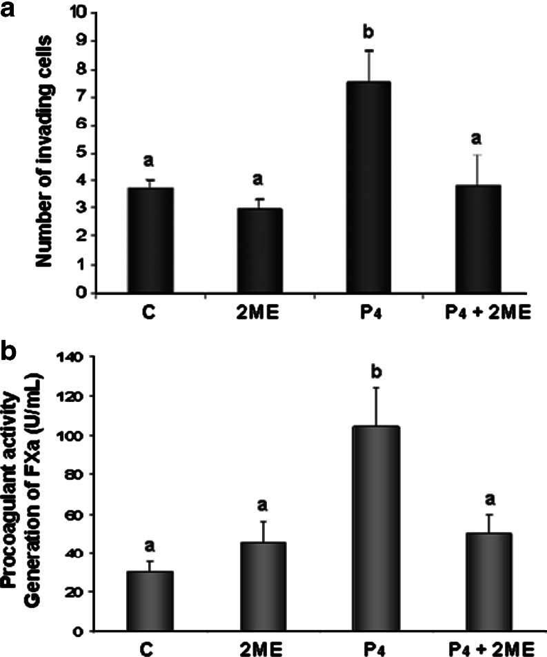
2-Methoxyestradiol (2ME) is an endogenous metabolite of 17β-estradiol with antiangiogenic and antitumor properties, although its mechanisms of action remain unclear. Progestins in hormone replacement therapy increase the risk of breast cancer. Progesterone also enhances the procoagulant activity and invasive potential of progesterone receptor (PR)-positive breast cancer cell lines, an effect largely mediated by induction of tissue factor (TF), the cellular activator of the coagulation cascade. Here we show that 2ME abrogates the induction TF expression in progesterone-treated breast cancer cells via a mechanism that does not involve the estrogen receptor. Instead, we demonstrate that by selectively antagonizing ERK1/2 signaling in breast cancer cells, 2ME limits the transactivation potential of ligand-bound PR and inhibits the expression of endogenous progesterone targets, such as TF and signal transducer and activator of transcription 5. We further demonstrate that 2ME can alter the phosphorylation status of PR. Thus, 2ME prevents progesterone-dependent increase in breast cancer cell invasiveness and procoagulant activity by uncoupling PR from the ERK1/2 signal transduction pathway.


Conflict of interest statement

The authors declare that they have no conflict of interest.

Figures

Fig. 1
Fig. 1
2ME inhibits induction of TF expression upon progesterone treatment. (a) ZR-75 breast cancer cells were treated with ethanol vehicle (C), 10 nM progesterone (P4), and increasing concentrations (1-10 µM) of 2ME alone or in combination for 24 hours before harvesting and TF quantification by Western Blotting (see methods). (b) Representative Western blot of TF and β-actin as in panel a except 2ME was applied at a fixed concentration of 5 µM for 24 hours. (c) ZR-75 breast cancer cells were treated with ethanol vehicle (C), 10 nM progesterone (P4), and 2ME (5 µM) alone or in combination for 24 hours before harvesting and TF mRNA evaluation by RT-PCR (Representative gel shown, see methods). The lower panels correspond to densitometric analysis in respect to β-actin and GAPDH for panels b and c respectively. Results are presented as mean ±SEM (minimum n=4).
Fig. 2
Fig. 2
2ME inhibits progesterone-induced invasion and procoagulation. a ZR-75 breast cancer cells were treated with ethanol vehicle (C), 10 nM progesterone (P4) and 5 μM 2ME alone in combination (P4 + 2ME) for 24 h before harvesting and quantification for invasion through a matrigel-coated porous membrane (see “Materials and Methods”). In each experiment, 15 fields (×200) were counted (n = 4 in duplicate). b ZR-75 cells were treated as in panel a before the quantification of procoagulant activity, as measured by the factor Xa generation (n = 8 in quintuplicate). The results are presented as mean±SEM.
Fig. 3
Fig. 3
Inhibition of progesterone responses by 2ME oocurs at the promoter level and does not require ER activation. a ZR-75 breast cancer cells were transfected with a luciferase fused to the human TF promoter construct and incubated with ethanol vehicle (C), 10 nM progesterone (P4) and 5 μM 2ME alone in combination (P4 + 2ME) for 20 h. Luciferase activity was normalized against activity from a cotranfected β-galactosidase vector. Results are mean±SEM of five different experiments, in triplicate (a ≠ b: P < 0.05). b TF expression in ZR-75 cells was assessed by Western blot analysis 24 h after treatment with 10 nM progesterone (P4) and/or 5 μM 2ME, in the presence or absence of ICI 182780 (ICI).
Fig. 4
Fig. 4
2ME inhibits progesterone mediated TF regulation in the T47D breast cancer cell line. a ER-negative T47D derived cells were treated with ethanol vehicle (C), 10 nM progesterone (P4), 5 μM 2ME, alone or in combination, for 6 h before PCR analysis of TF mRNA expression. Glyceraldehyde-3-phosphate dehydrogenase mRNA levels were used as an internal control. b The T47D derived cell line was treated as in panel a for 24 h and TF protein levels determined by Western blotting. Representative results of three independent experiments are shown. c The procoagulant activity, quantified by the generation of factor Xa, was measured in T47D cells treated as in panel a. The results represent the mean procoagulant activity (±SEM) of three independent experiments, each measured in quintuplicate (a ≠ b: P < 0.05).
Fig. 5
Fig. 5
2ME reduces both basal and progesterone-increased ERK1/2 and c-RAF phosphorylation. a ZR-75 cells in serum-free medium were pretreated with ethanol vehicle or 10 nM progesterone (P4) for 18 h before the addition of 5 μM 2ME for 5, 15 and 30 min, before Western blotting for levels of total (t-) and phosphorylated (p-) ERK1 and ERK2 (ERK1/2). The lower panel represents the densitometric analysis of phosphorylated ERK1 and ERK2 normalized to total ERK1 and ERK2 in three independent experiments (n = 3). Statistical analysis was performed using Kluskal–Wallis with Dunn post-test methods (a ≠ b, P < 0.05). b Western blots of phosphorylated AKT and c-RAF in cells pretreated with ethanol vehicle or 10 nM progesterone (P4) for 18 h before the addition of 5 μM 2ME for 5, 15 and 30 min.
Fig. 6
Fig. 6
Progesterone requires the MAPK pathway to increase TF protein and coagulation. a ZR-75 breast cancer cells were treated with vehicle (ethanol and DMSO) or 10 nM progesterone (P4) in the presence of MEK-1 inhibitor (PD, PD98059), and the AKT and PI3-K inhibitors [AKTX and wortmannin (WORT), respectively] for 24 h, before harvesting and TF quantification by Western blotting (see “Materials and Methods”) (n = 3). n.s. nonspecific band. b The subsequent requirement of MEK1 for coagulation in the presence of progesterone was also confirmed by the quantification of procoagulant activity, as measured by the factor Xa generation. The results are presented as mean±SEM, n = 4 in quintuplicate (a ≠ b: P < 0.05).
Fig. 7
Fig. 7
2ME inhibits progesterone-increased PR phosphorylation at serine 400. a ZR-75 cells in serum-free medium were treated with ethanol vehicle (C) or 10 nM progesterone (P4) with or without 5 μM 2ME for 15 and 30 min before Western blot analysis of PR serine 400 phosphorylation (p-S400-PRB). The lower panel depicts densitometric analysis of the observed changes of p-S400-PRB normalized to total PR-B. The results are presented as mean±SEM, n = 3 (a ≠ b; c ≠ d, P < 0.05).
Fig. 8
Fig. 8
2ME inhibits other progesterone-regulated genes: partial reduction off a canonical PRE and inhibition of STAT5. a ZR-75 cells were transfected with a PRE-Luc reporter construct and treated with ethanol vehicle (C), 10 nM progesterone (P4), 5 μM 2ME alone or in combination for 20 h before harvesting and luciferase quantification against β-galactosidase (see “Materials and Methods”). b ZR-75 cells were treated as in panel a for 24 h before harvesting and STAT5a and STAT5b Western blot analysis (see “Materials and Methods”). c ZR-75 breast cancer cells were as in panel a for 6 h before harvesting and STAT5b mRNA quantification by PCR (see “Materials and Methods”). d ZR-75 cells were treated with vehicle (ethanol and DMSO; C) or 10 nM progesterone (P4) in the presence or absence of MEK-1, AKT and PI3-K inhibitors (PD, AKTX and WORT, respectively) for 24 h, before harvesting and TF analysis by Western blotting (representative blot of three independent experiments). (a ≠ b, b ≠ c, c ≠ a, P < 0.05).

References

    1. Zhu B, Conney AH. Functional role of estrogen metabolism in target cells: review and perspectives. Carcinogenesis. 1998;19(1):1–27. doi: 10.1093/carcin/19.1.1. - DOI - PubMed
    1. Muti P, Bradlow HL, Micheli A, Krogh V, Freudenheim JL, Schünemann HJ, Stanulla M, Yang J, Sepkovic DW, Trevisan M, Berrino F. Estrogen metabolism and risk of breast cancer: a prospective study of the 2:16alpha-hydroxyestrone ratio in premenopausal and postmenopausal women. Epidemiology. 2000;11(6):635–640. doi: 10.1097/00001648-200011000-00004. - DOI - PubMed
    1. Van Zijl C, Lottering ML, Steffens F, Joubert A. In vitro effects of 2-methoxyestradiol on MCF-12A and MCF-7 cell growth, morphology and mitotic spindle formation. Cell Biochem Funct. 2008;26(5):632–642. doi: 10.1002/cbf.1489. - DOI - PubMed
    1. Mooberry SL. New insights into 2-methoxyestradiol, a promising antiangiogenic and antitumor agent. Curr Opin Oncol. 2003;15(6):425–430. doi: 10.1097/00001622-200311000-00004. - DOI - PubMed
    1. Dahut WL, Lakhani NJ, Gulley JL, Arlen PM, Kohn EC, Kotz H, McNally D, Parr A, Nguyen D, Yang SX, Steinberg SM, Venitz J, Sparreboom A, Figg WD. Phase I clinical trial of oral 2-methoxyestradiol, an antiangiogenic and apoptotic agent, in patients with solid tumors. Cancer Biol Ther. 2006;5(1):22–27. doi: 10.4161/cbt.5.1.2349. - DOI - PubMed

Publication types

MeSH terms