

Adiponectin receptor expression in human malignant tissues
- PMID: 21761356
- PMCID: PMC10358112
- DOI: 10.1007/s12672-010-0017-7
Adiponectin receptor expression in human malignant tissues
Abstract
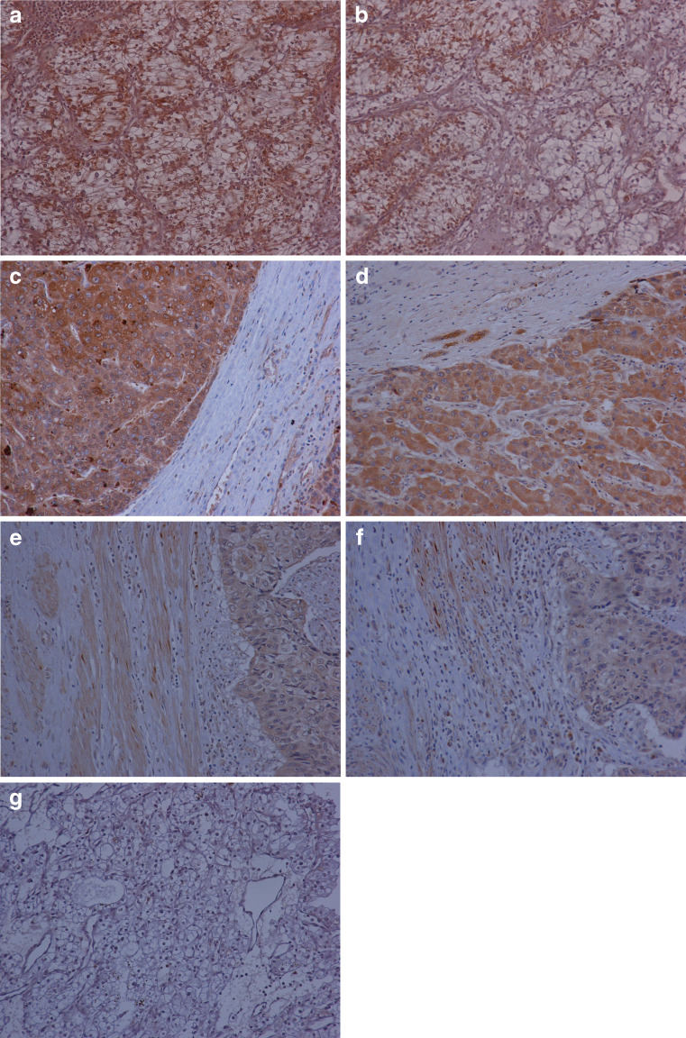
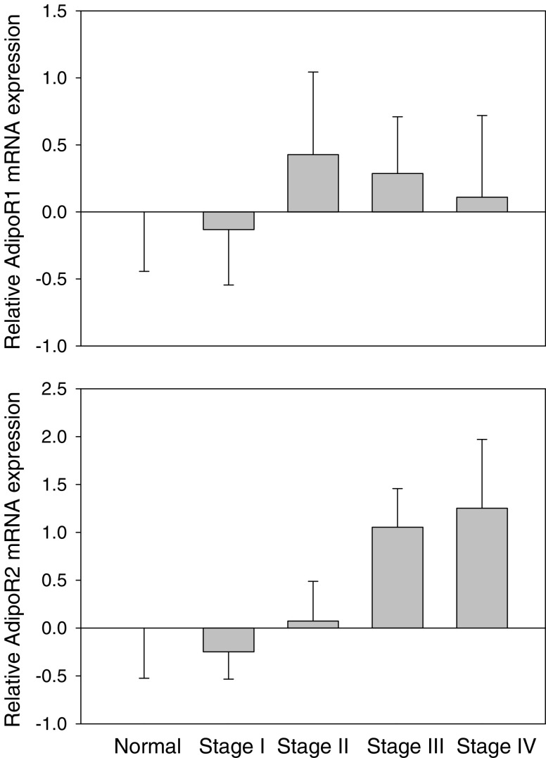
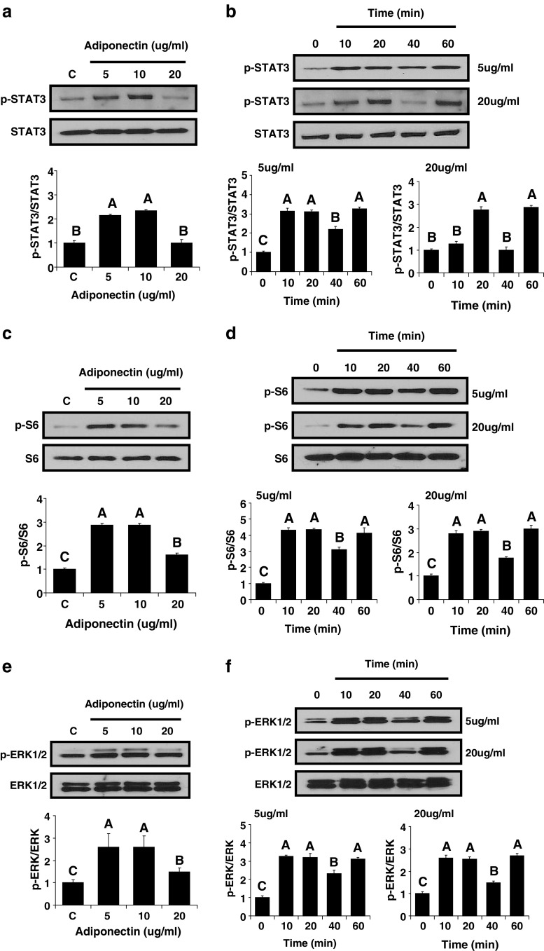
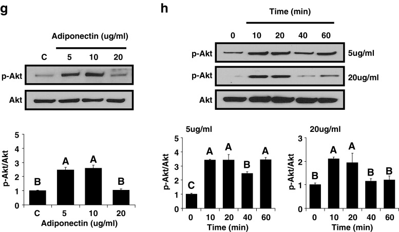
Adiponectin has been proposed to be a mediator of obesity-associated malignancies and to have direct antineoplastic effects acting via adiponectin receptors AdipoR1 and AdipoR2. We describe herein the expression of AdipoR1 and AdipoR2 in several cancers not previously studied. We used immunohistochemistry to assess expression of adiponectin receptors in archival specimens of renal cell carcinoma (n = 64), hepatocellular carcinoma (n = 123), melanoma (n = 20), cholangiocarcinoma (n = 20), transitional cell carcinoma of the bladder (n = 24), ovarian epithelial carcinoma (n = 63), cervical squamous cell carcinoma (n = 49), and adrenocortical carcinoma (n = 48). To compare expression in malignant versus nonmalignant tissues, we also studied AdipoR1 and AdipoR2 expression in pairs of renal cell carcinoma and adjacent healthy kidney tissue specimens by immunohistochemistry. We also studied mRNA expression in 45 specimens of renal cell carcinoma by real-time polymerase chain reaction. Finally, we utilized Western blotting to confirm the presence of adiponectin receptors and subsequently studied cell signaling pathways of adiponectin in the renal cancer cell line 786-O. Cancers associated with obesity were significantly more likely to express AdipoR1 than cancers not associated with obesity. Of the specimens of renal cell carcinoma, which is strongly associated with obesity, 93.8% expressed AdipoR1 compared to 44.9% of the specimens of cervical cell carcinoma, which is not associated with obesity (p < 0.001). There was no difference in the expression of adiponectin receptors or their mRNA between malignant and benign kidney tissue specimens. Overall, there were no correlations between expression of adiponectin receptors or their mRNA and tumor prognostic factors. Finally, Western blotting confirmed the presence of AdipoR1 in the renal cancer cell line 786-O, and adiponectin activates in vitro several signaling pathways in this cell line. In summary, we report for the first time expression of AdipoR1 and AdipoR2 in the above cancers and that AdipoR1 is more ubiquitously expressed in obesity-associated cancers.
Figures
References
-
- Barb D, Williams CJ, Neuwirth AK, et al. Adiponectin in relation to malignancies: a review of existing basic research and clinical evidence. Am J Clin Nutr. 2007;86:s858–s866. - PubMed
-
- Guadagni F, Roselli M, Martini F, et al. Prognostic significance of serum adipokine levels in colorectal cancer patients. Anticancer Res. 2009;29:3321–3327. - PubMed
MeSH terms
Substances
LinkOut - more resources
Full Text Sources
Other Literature Sources
