. 2012 Mar;18(3):460-71.
doi: 10.1002/ibd.21812. Epub 2011 Jul 14.

Intestinal fibrosis is reduced by early elimination of inflammation in a mouse model of IBD: impact of a "Top-Down" approach to intestinal fibrosis in mice

Affiliations

Intestinal fibrosis is reduced by early elimination of inflammation in a mouse model of IBD: impact of a "Top-Down" approach to intestinal fibrosis in mice

Laura A Johnson et al. Inflamm Bowel Dis. 2012 Mar.

Abstract

Background: The natural history of Crohn's disease follows a path of progression from an inflammatory to a fibrostenosing disease, with most patients requiring surgical resection of fibrotic strictures. Potent antiinflammatory therapies reduce inflammation but do not appear to alter the natural history of intestinal fibrosis. The aim of this study was to determine the relationship between intestinal inflammation and fibrogenesis and the impact of a very early "top-down" interventional approach on fibrosis in vivo.

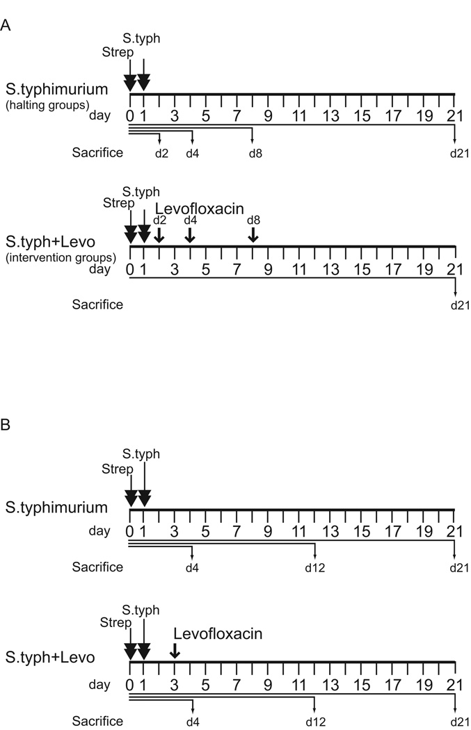
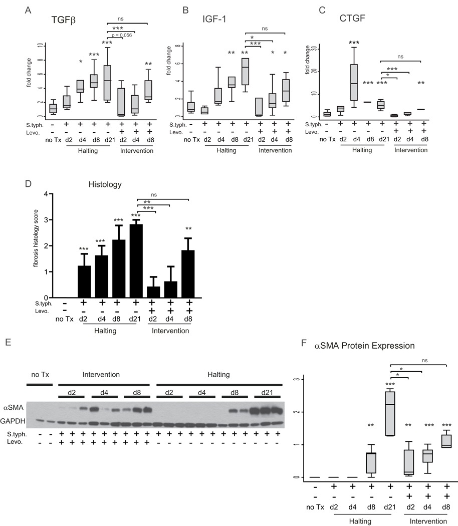
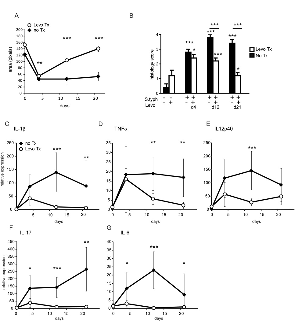
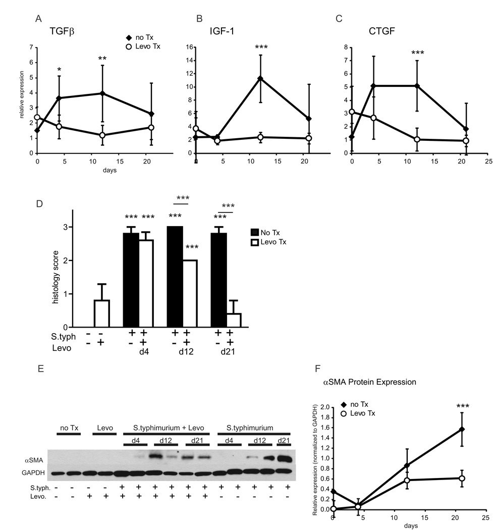
Methods: In this study we removed the inflammatory stimulus from the Salmonella typhimurium mouse model of intestinal fibrosis by eradicating the S. typhimurium infection with levofloxacin at sequential timepoints during the infection. We evaluated the effect of this elimination of the inflammatory stimulus on the natural history of inflammation and fibrosis as determined by gross pathology, histopathology, mRNA expression, and protein expression.

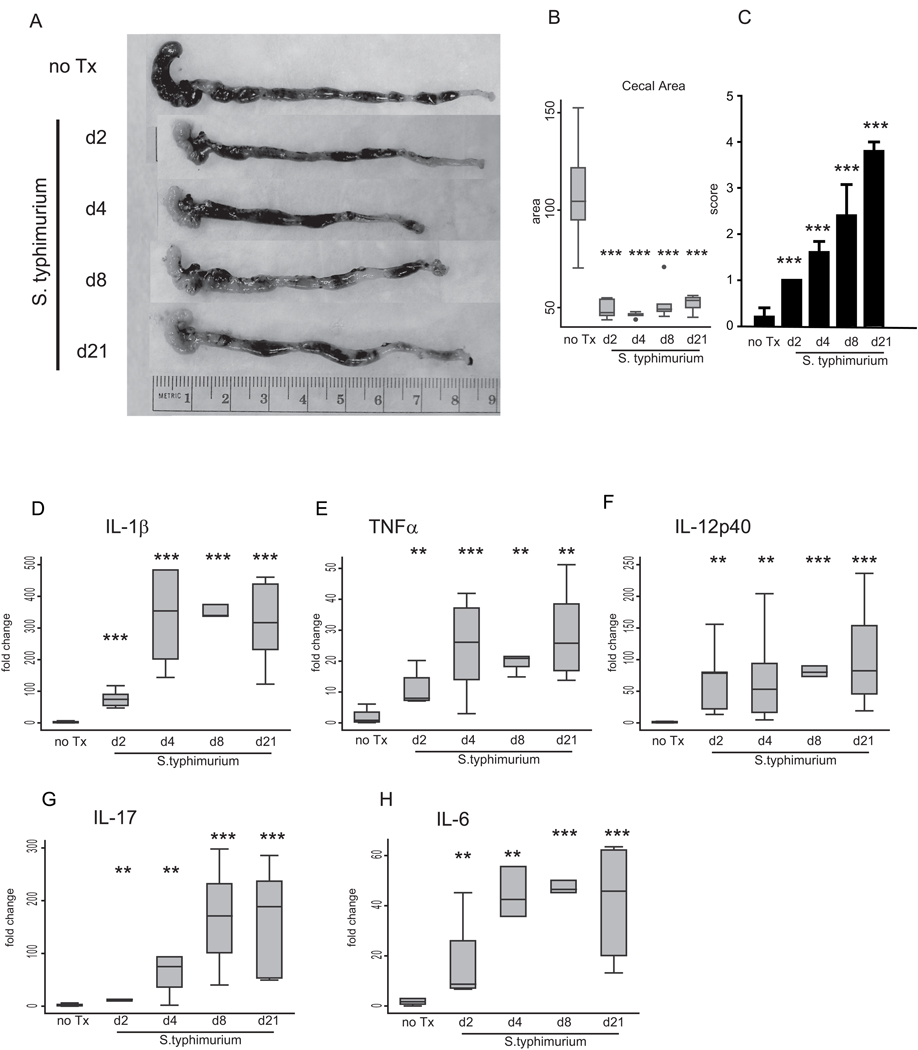
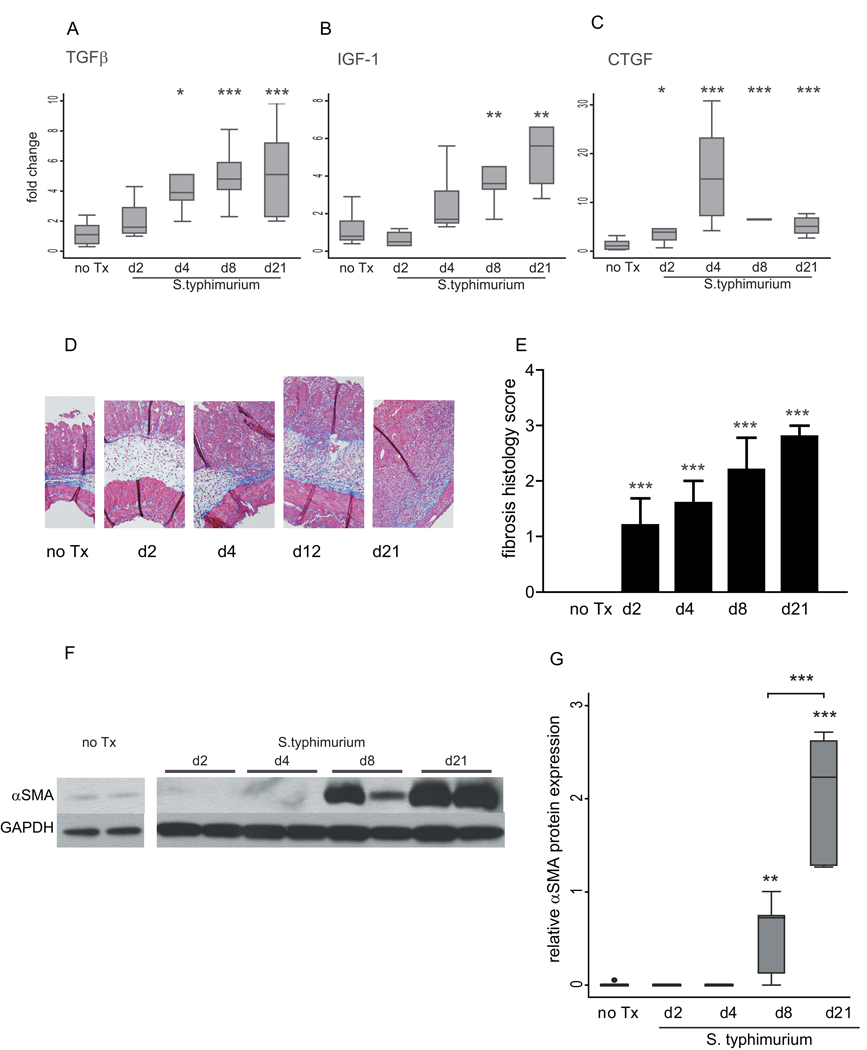
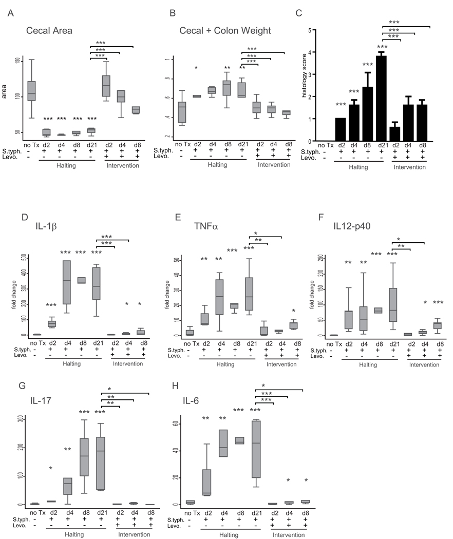
Results: Fibrogenesis is preceded by inflammation. Delayed eradication of the inflammatory stimulus by antibiotic treatment represses inflammation without preventing fibrosis. Early intervention significantly ameliorates but does not completely prevent subsequent fibrosis.

Conclusions: This study demonstrates that intestinal fibrosis develops despite removal of an inflammatory stimulus and elimination of inflammation. Early intervention ameliorates but does not abolish subsequent fibrosis, suggesting that fibrosis, once initiated, is self-propagating, suggesting that a very early top-down interventional approach may have the most impact on fibrostenosing disease.


Figures

Figure 1
Figure 1. Treatment schematic of levofloxacin endpoint analysis and time course comparison experiments
(A) Treatment schematic of the endpoint analysis experiment. Mice in the S. typhimurium infection halting groups (upper panel) were treated with streptomycin (strep, double arrows) 1 day prior to infection with S. typhimurium (S. typh, double arrows) and sacrificed at day 2, 4, 8 and 21 post-infection. In the levofloxacin intervention groups (lower panel), mice received streptomycin and S. typhimurium on the same schedule. Levofloxacin intervention (single arrows) began day 2, 4, or 8 post-S. typhimurium infection and continued until sacrifice at day 21 post-infection. (B) Treatment schematic of the time course comparison experiments. Mice in the S. typhimurium infection groups (upper panel) were treated with streptomycin (strep, double arrows) 1 day prior to infection with S. typhimurium (S.typh, double arrows) and sacrificed day 4, 12 and 21 post-infection. In the levofloxacin intervention time course (lower panel) mice received streptomycin and S. typhimurium on the same schedule. Levofloxacin intervention (arrow) began day 3 post-S. typhimurium infection and continued until sacrifice at day 4, 12, and 21 post-infection. In all experiments, 5 animals were used per experimental group.
Figure 2
Figure 2. Inflammation precedes fibrosis in the S. typhimurium mouse model of intestinal fibrosis
(A) Representative gross appearance of the cecum and distal colon of S. typhimurium-infected mice day 2 (d2), day 4 (d4), day 8 (d8) and day 21 (d21) post-infection compared to uninfected (no Tx) against a 1-cm reference ruler. (B) Cecal area calculated from uninfected mice (no Tx) compared to S. typhimurium-infected mice day 2, 4, 8, and 21 post-infection. (C) Blinded inflammatory scoring of cecal sections using the 0–4 point Wirtz scale. (D–H) qRT-PCR gene expression of Th1 and inflammatory cytokines in the cecum of uninfected (no Tx) compared to S. typhimurium-infected mice day 2, 4, 8, and 21 post-infection. Gene expression was normalized to GAPDH expression. Statistical comparisons between uninfected controls (no Tx) and the infected groups are denoted with asterisks. * p <0.05, ** p<0.01, *** p<0.001
Figure 3
Figure 3. Development of fibrosis in the S. typhimurium mouse model of fibrosis
(A–C) qRT-PCR expression of fibrotic genes in cecum from uninfected mice (no Tx) compared to S. typhimurium-infected mice day 2, 4, 8, and 21 post-infection. Gene expression was normalized to GAPDH expression. (D) Representative trichrome histological sections (100× magnification). (E) Blinded fibrosis scoring of cecal sections was determined from trichrome stained cecal sections. (F) Representative Western blot of αSMA protein expression in S. typhimurium mice days 2, 4, 8, and 21 post-infection compared to uninfected controls. The samples shown were run on the same protein gel. The image has been cut to rearrange the loading order for clarity. GAPDH expression was used as a loading control. (G) Quantitation of αSMA protein expression in cecum from uninfected mice (no Tx) compared to S. typhimurium infected mice day 2, 4, 8, and 21 post-infection. Western blots were digitally scanned and quantitated using ImageJ. Relative expression was normalized to GAPDH protein expression. Statistical comparisons between uninfected controls (no Tx) and the infected groups are denoted with asterisks. Brackets represent statistical comparisons between S. typhimurium-infected groups. * p <0.05, ** p<0.01, *** p<0.001
Figure 4
Figure 4. Intervention represses the inflammatory response
(A) Cecal area from uninfected mice (no Tx) compared to S. typhimurium-infected mice day 2, 4, 8, and 21 post-infection, and S. typhimurium-infected mice treated with levofloxacin day 2, 4, and 8 post-infection and sacrificed day 21 post-infection. (B) Combined cecal + colon weight. (C) Blinded inflammatory scoring of cecal sections using the 0–4 point Wirtz scale. (D–H) qRT-PCR expression of IL-1β (D), TNFα (E), IL-12p40 (F), IL-17 (G), and IL-6 (H) in mice infected with S. typhimurium-infected mice day 2, 4, 8, and 21 post-infection, and S. typhimurium-infected mice treated with levofloxacin day 2, 4, and 8 post-infection and sacrificed day 21 post-infection compared to uninfected mice (no Tx). Statistical comparisons between the infection groups or intervention groups and the uninfected controls are denoted with asterisks. Statistically significant comparisons between the S. typhimurium infected and levofloxacin intervention groups at day 21 post-infection are denoted in brackets. * p <0.05, ** p<0.01, *** p<0.001
Figure 5
Figure 5. Early intervention ameliorates but does not completely prevent fibrosis
(A–C) qRT-PCR expression of fibrotic markers TGFβ (A), IGF-1 (B), and CTGF (C) in mice infected with S. typhimurium day 2, 4, 8, and 21 post-infection, and S. typhimurium-infected mice treated with levofloxacin day 2, 4, and 8 post-infection and sacrificed day 21 post-infection compared to uninfected mice (no Tx). (D) Blinded fibrosis scoring of cecal sections was determined from trichrome stained cecal sections. (E) Representative Western blot illustrating αSMA protein expression in the cecum of uninfected mice (no Tx) compared to mice infected with S. typhimurium and treated with levofloxacin 2, 4, and 8 post-infection and sacrificed day 21 post-infection and mice infected with S. typhimurium-infected mice day 2, 4, 8, and 21 post-infection. GAPDH protein expression was used as a loading control. (F) Quantitation of αSMA protein expression in the cecum. Western blots were digitally scanned and quantitated using ImageJ. Relative expression was normalized to GAPDH protein expression. Statistical comparisons between the infection groups or intervention groups and the uninfected controls are denoted with asterisks. Statistically significant comparisons between the S. typhimurium-infected and levofloxacin intervention groups at day 21 post-infection are denoted in brackets. ns = not statistically significant, * p <0.05, ** p<0.01, *** p<0.001
Figure 6
Figure 6. Early intervention represses inflammation
(A) Levofloxacin intervention reverses cecal contraction in response to S. typhimurium infection. Cecal area of mice infected with S. typhimurium (solid diamonds) compared to S. typhimurium-infected animals treated with levofloxacin day 3 post-infection (open circles) at day 4, 12, and 21 post-infection. (B) Blinded inflammatory scoring of cecal sections using the 0–4 point Wirtz scale. (C–G) qRT-PCR expression of IL-1β, TNFα IL-12p40, IL-17, and IL-6 in the cecum day 4, 12, and 21 post-infection. Statistical comparisons between the infection and intervention groups are denoted with asterisks. *p <0.05, ** p<0.01, *** p<0.001
Figure 7
Figure 7. Early intervention represses inflammation but does not prevent fibrosis
(A–C) qRT-PCR expression of fibrotic markers TGFβ (A), IGF-1 (B), and CTGF (C) in mice infected with S. typhimurium (solid diamonds) compared to S. typhimurium-infected mice treated with levofloxacin day 3 post-infection (open circles) day 4, 12, and 21 post-infection. (D) Blinded fibrosis scoring of cecal sections was determined from trichrome stained cecal sections. (E) Representative Western blot of cecal αSMA protein expression in mice infected with S. typhimurium compared to mice infected with S. typhimurium and treated with levofloxacin, uninfected mice (no Tx), and levofloxacin treated uninfected mice (Levo). GAPDH was used as a control for protein loading. (F) αSMA protein expression in the cecum in response to S. typhimurium infection or S. typhimurium infection and levofloxacin intervention. Relative αSMA expression was normalized to GAPDH expression. Statistically significant comparisons between the infection and intervention groups are denoted with asterisks. * p <0.05, ** p<0.01, *** p<0.001

References

    1. Andres PG, Friedman LS. Epidemiology and the natural course of inflammatory bowel disease. Gastroenterol Clin North Am. 1999;28:255–281. vii. - PubMed
    1. Sands BE, Arsenault JE, Rosen MJ, et al. Risk of early surgery for Crohn's disease: implications for early treatment strategies. Am J Gastroenterol. 2003;98:2712–2718. - PubMed
    1. Cosnes J, Cattan S, Blain A, et al. Long-term evolution of disease behavior of Crohn's disease. Inflamm Bowel Dis. 2002;8:244–250. - PubMed
    1. Feagan BG, Vreeland MG, Larson LR, et al. Annual cost of care for Crohn's disease: a payor perspective. Am J Gastroenterol. 2000;95:1955–1960. - PubMed
    1. Lichtenstein GR, Olson A, Travers S, et al. Factors associated with the development of intestinal strictures or obstructions in patients with Crohn's disease. Am J Gastroenterol. 2006;101:1030–1038. - PubMed

Publication types

MeSH terms