

Segmentation of brain images using adaptive atlases with application to ventriculomegaly
- PMID: 21761641
- PMCID: PMC3478153
- DOI: 10.1007/978-3-642-22092-0_1
Segmentation of brain images using adaptive atlases with application to ventriculomegaly
Abstract
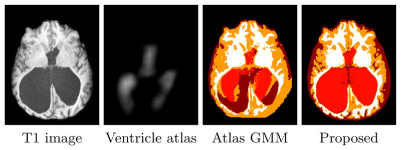
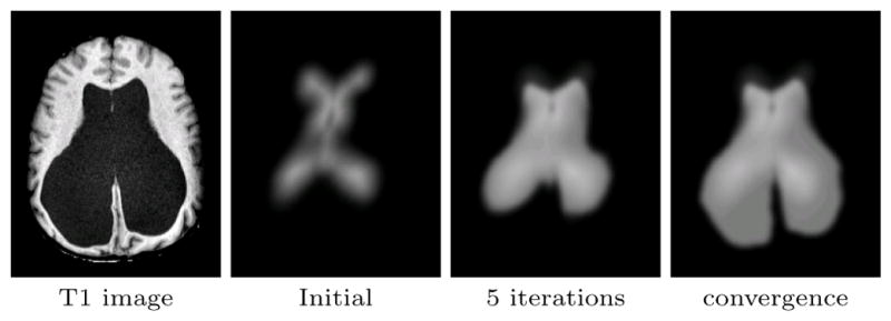
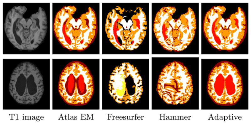
Segmentation of brain images often requires a statistical atlas for providing prior information about the spatial position of different structures. A major limitation of atlas-based segmentation algorithms is their deficiency in analyzing brains that have a large deviation from the population used in the construction of the atlas. We present an expectation-maximization framework based on a Dirichlet distribution to adapt a statistical atlas to the underlying subject. Our model combines anatomical priors with the subject's own anatomy, resulting in a subject specific atlas which we call an "adaptive atlas". The generation of this adaptive atlas does not require the subject to have an anatomy similar to that of the atlas population, nor does it rely on the availability of an ensemble of similar images. The proposed method shows a significant improvement over current segmentation approaches when applied to subjects with severe ventriculomegaly, where the anatomy deviates significantly from the atlas population. Furthermore, high levels of accuracy are maintained when the method is applied to subjects with healthy anatomy.
Figures
References
-
- Ashburner J, Friston K. Multimodal image coregistration and partitioning–a unified framework. NeuroImage. 1997;6(3):209–17. - PubMed
-
- Bhatia KK, Aljabar P, Boardman JP, Srinivasan L, Murgasova M, Counsell SJ, Rutherford MA, Hajnal J, Edwards AD, Rueckert D. Groupwise combined segmentation and registration for atlas construction. In: Ayache N, Ourselin S, Maeder A, editors. Proc of MICCAI. pp. 532–40. - PubMed
- Lecture Notes in Computer Science. Springer; Berlin / Heidelberg: 2007.
-
- Clarke MJ, Meyer FB. The history of mathematical modeling in hydrocephalus. Neurosurg Focus. 2007;22(4):E3. - PubMed
-
- Collins DL, Zijdenbos AP, Kollokian V, Sled JG, Kabani NJ, Holmes CJ, Evans AC. Design and construction of a realistic digital brain phantom. IEEE Trans Med Imaging. 1998;17(3):463–468. - PubMed
MeSH terms
Grants and funding
LinkOut - more resources
Full Text Sources
Medical
Miscellaneous