Feasibility of a femoral nerve motor branch for transfer to the pudendal nerve for restoring continence: a cadaveric study
- PMID: 21761966
- PMCID: PMC3277789
- DOI: 10.3171/2011.6.SPINE11163
Feasibility of a femoral nerve motor branch for transfer to the pudendal nerve for restoring continence: a cadaveric study
Abstract
Object: Nerve transfers are an effective means of restoring control to paralyzed somatic muscle groups and, recently, even denervated detrusor muscle. The authors performed a cadaveric pilot project to examine the feasibility of restoring control to the urethral and anal sphincters using a femoral motor nerve branch to reinnervate the pudendal nerve through a perineal approach.
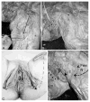
Methods: Eleven cadavers were dissected bilaterally to expose the pudendal and femoral nerve branches. Pertinent landmarks and distances that could be used to locate these nerves were assessed and measured, as were nerve cross-sectional areas.
Results: A long motor branch of the femoral nerve was followed into the distal vastus medialis muscle for a distance of 17.4 ± 0.8 cm, split off from the main femoral nerve trunk, and transferred medially and superiorly to the pudendal nerve in the Alcock canal, a distance of 13.7 ± 0.71 cm. This was performed via a perineal approach. The cross-sectional area of the pudendal nerve was 5.64 ± 0.49 mm(2), and the femoral nerve motor branch at the suggested transection site was 4.40 ± 0.41 mm(2).
Conclusions: The use of a femoral nerve motor branch to the vastus medialis muscle for heterotopic nerve transfer to the pudendal nerve is surgically feasible, based on anatomical location and cross-sectional areas.
Figures


Similar articles
-
Ipsilateral S2 nerve root transfer to pudendal nerve for restoration of external anal and urethral sphincter function: an anatomical study.Sci Rep. 2019 Sep 30;9(1):13993. doi: 10.1038/s41598-019-50484-7. Sci Rep. 2019. PMID: 31570751 Free PMC article.
-
Reinnervation of urethral and anal sphincters with femoral motor nerve to pudendal nerve transfer.Neurourol Urodyn. 2011 Nov;30(8):1695-704. doi: 10.1002/nau.21171. Epub 2011 Sep 26. Neurourol Urodyn. 2011. PMID: 21953679 Free PMC article.
-
Anatomical feasibility of performing a nerve transfer from the femoral branch to bilateral pelvic nerves in a cadaver: a potential method to restore bladder function following proximal spinal cord injury.J Neurosurg Spine. 2013 Jun;18(6):598-605. doi: 10.3171/2013.2.SPINE12793. Epub 2013 Mar 29. J Neurosurg Spine. 2013. PMID: 23540734 Free PMC article.
-
Neuronal innervation of urethral and anal sphincters: surgical anatomy and clinical implications.Curr Opin Obstet Gynecol. 2000 Oct;12(5):387-98. doi: 10.1097/00001703-200010000-00008. Curr Opin Obstet Gynecol. 2000. PMID: 11111881 Review.
-
Current status: new technologies for the treatment of patients with fecal incontinence.Surg Endosc. 2014 Aug;28(8):2277-301. doi: 10.1007/s00464-014-3464-3. Epub 2014 Mar 8. Surg Endosc. 2014. PMID: 24609699 Review.
Cited by
-
Bladder reinnervation using a primarily motor donor nerve (femoral nerve branches) is functionally superior to using a primarily sensory donor nerve (genitofemoral nerve).J Urol. 2015 Mar;193(3):1042-51. doi: 10.1016/j.juro.2014.07.095. Epub 2014 Jul 24. J Urol. 2015. PMID: 25066874 Free PMC article.
-
Ipsilateral S2 nerve root transfer to pudendal nerve for restoration of external anal and urethral sphincter function: an anatomical study.Sci Rep. 2019 Sep 30;9(1):13993. doi: 10.1038/s41598-019-50484-7. Sci Rep. 2019. PMID: 31570751 Free PMC article.
-
Sciatic Nerve to Pudendal Nerve Transfer: Anatomical Feasibility for a New Proposed Technique.Indian J Plast Surg. 2019 May;52(2):222-225. doi: 10.1055/s-0039-1688513. Epub 2019 May 6. Indian J Plast Surg. 2019. PMID: 31602139 Free PMC article.
-
Nerve transfer for restoration of lower motor neuron-lesioned bladder and urethra function: establishment of a canine model and interim pilot study results.J Neurosurg Spine. 2019 Nov 8;32(2):258-268. doi: 10.3171/2019.8.SPINE19265. Print 2020 Feb 1. J Neurosurg Spine. 2019. PMID: 31703192 Free PMC article.
-
Nerve transfer for restoration of lower motor neuron-lesioned bladder function. Part 2: correlation between histological changes and nerve evoked contractions.Am J Physiol Regul Integr Comp Physiol. 2021 Jun 1;320(6):R897-R915. doi: 10.1152/ajpregu.00300.2020. Epub 2021 Mar 24. Am J Physiol Regul Integr Comp Physiol. 2021. PMID: 33759573 Free PMC article.
References
-
- Anderson KD. Targeting recovery: priorities of the spinal cord-injured population. J Neurotrauma. 2004;21:1371–1383. - PubMed
-
- Boger A, Bhadra N, Gustafson KJ. Bladder voiding by combined high frequency electrical pudendal nerve block and sacral root stimulation. Neurourol Urodyn. 2008;27:435–439. - PubMed
-
- Browne EZ, Snyder CC. Intercosto-sacral neural anastomosis. Surg Forum. 1971;22:474–476. - PubMed
-
- Carlsson CA, Sundin T. Reconstruction of efferent pathways to the urinary bladder in a paraplegic child. Rev Surg. 1967;24:73–76. - PubMed
Publication types
MeSH terms
Grants and funding
LinkOut - more resources
Full Text Sources
Medical

