. 2011 Jul 15:9:1-12.
doi: 10.1186/1479-5876-9-111.

Complement component C5a promotes expression of IL-22 and IL-17 from human T cells and its implication in age-related macular degeneration

Affiliations

Complement component C5a promotes expression of IL-22 and IL-17 from human T cells and its implication in age-related macular degeneration

Baoying Liu et al. J Transl Med. .

Abstract

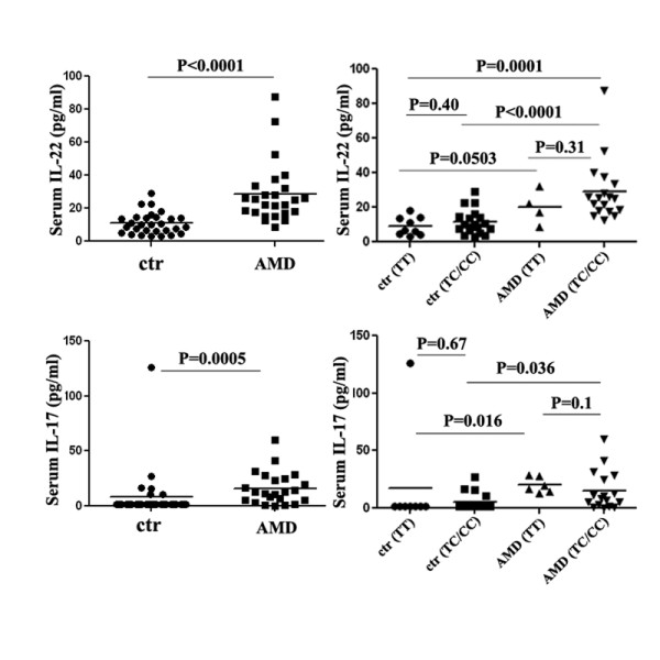
Background: Age related macular degeneration (AMD) is the leading cause of irreversible blindness in elderly populations worldwide. Inflammation, among many factors, has been suggested to play an important role in AMD pathogenesis. Recent studies have demonstrated a strong genetic association between AMD and complement factor H (CFH), the down-regulatory factor of complement activation. Elevated levels of complement activating molecules including complement component 5a (C5a) have been found in the serum of AMD patients. Our aim is to study whether C5a can impact human T cells and its implication in AMD.

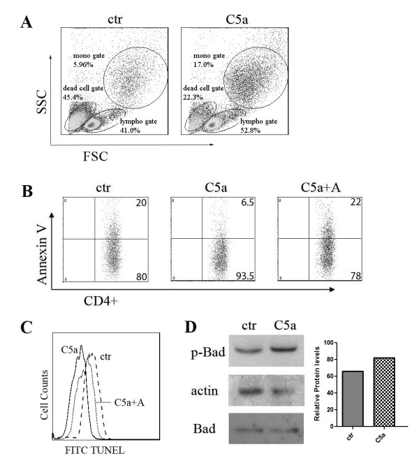
Methods: Human peripheral blood mononuclear cells (PBMCs) were isolated from the blood of exudative form of AMD patients using a Ficoll gradient centrifugation protocol. Intracellular staining and enzyme-linked immunosorbent assays were used to measure protein expression. Apoptotic cells were detected by staining of cells with the annexin-V and TUNEL technology and analyzed by a FACS Caliber flow cytometer. SNP genotyping was analyzed by TaqMan genotyping assay using the Real-time PCR system 7500.

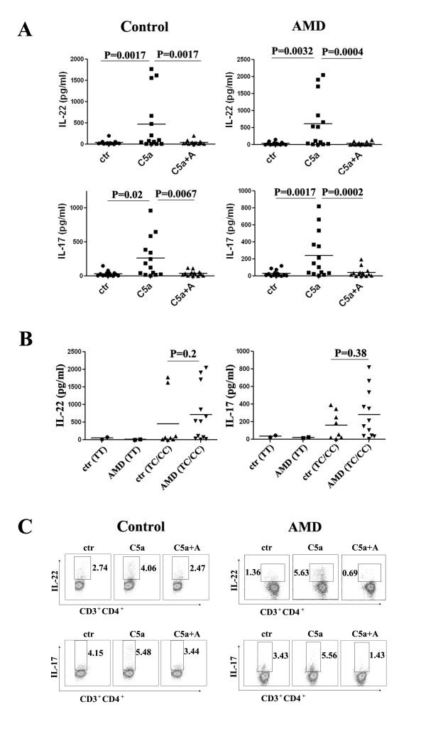
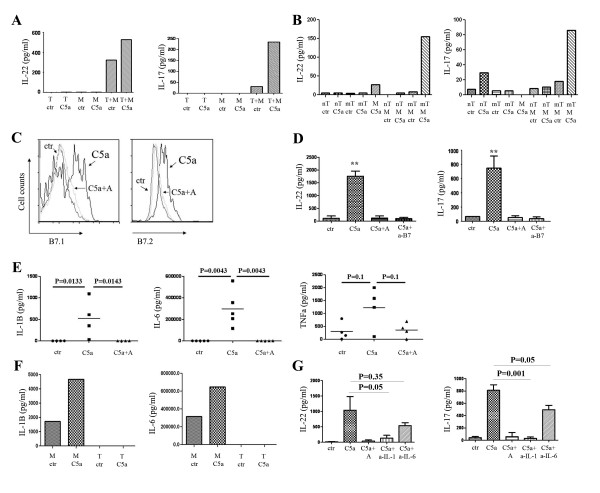
Results: We show that C5a promotes interleukin (IL)-22 and IL-17 expression by human CD4+ T cells. This effect is dependent on B7, IL-1β and IL-6 expression from monocytes. We have also found that C5a could protect human CD4+ cells from undergoing apoptosis. Importantly, consistent with a role of C5a in promoting IL-22 and IL-17 expression, significant elevation in IL-22 and IL-17 levels was found in AMD patients as compared to non-AMD controls.

Conclusions: Our results support the notion that C5a may be one of the factors contributing to the elevated serum IL-22 and IL-17 levels in AMD patients. The possible involvement of IL-22 and IL-17 in the inflammation that contributes to AMD may herald a new approach to treat AMD.


Figures

Figure 1
Figure 1
C5a promotes the expression of IL-22 and IL-17 from T cells. (A) IL-22 and IL-17 in 3-day culture supernatants of PBMCs from 14 AMD patients and 14 controls. (B) C5a induced IL-22/IL-17 expression in both controls and AMD patients were subgrouped based on CFH genotypes. (C) Intracytoplasmic staining of IL-22 and IL-17 from both controls and AMD patients after 5 days of culture with or without C5a and C5aR antagonist.
Figure 2
Figure 2
IL-1β and IL-6 secreting monocytes are important for C5a induced IL-22 and IL-17 expression form T cells. (A) CD3+CD4+ T (T) cells and CD3-CD14+ monocytes (M) were sorted and cultured with or without C5a for 3 days. Cell supernatants were assessed for IL-22 and IL-17 expression. Three separate experiments were performed and the figure shows representative data. (B) CD3+CD4+CD45RA+ (naïve T cells, nT) and CD3+CD4+CD45RA- (memory T cells, mT) T cells and CD3-CD14+ monocytes (M) were sorted and cultured with or without C5a for 3 days. IL-22 and IL-17 levels were measured from supernatants. Three separate experiments were performed and the figure shows representative data. (C) C5a activates B7 expression on monocytes. PBMCs were cultured with or without C5a for 1 day. CD3-CD14+ monocytes were gated for indicated cell markers' expression. Similar results were seen in another independent assay. (D) IL-22 and IL-17 in 3-day culture supernatants of PBMCs with the presence or absence of C5a, C5aR antagonist and anti-B7.1 and anti-B7.2 antibodies. (E) C5a stimulates monocytes to secrete IL-1β and IL-6. PBMCs were cultured with or without C5a and C5aR antagonist for 3 days. Cell supernatants were assayed for IL-1β, IL-6 and TNFα expression. (F) Monocytes and T cells were sorted and cultured with or without C5a for 3 day. Cell supernatants were assayed for IL-1β and IL-6 expression. Three separate experiments were performed and the figure shows representative data. (G) IL-22 and IL-17 in 3-day culture supernatants of PBMCs with the presence or absence of C5a with isotype control antibody, C5aR antagonist and anti-IL-1β and anti-IL-6 neutralization antibodies. Three separate experiments were performed.
Figure 3
Figure 3
C5a protects T cells from undergoing apoptosis. (A) Scatter plot of PBMCs cultured with or without C5a. Three separate experiments were performed and the figure shows representative data. (B) Annexin V expression on T cells cultured with or without C5a and C5aR antagonist. Ten separate experiments were performed and the figure shows representative data. (C) TUNEL staining of CD4+ T cells treated with or without C5a and C5aR antagonist. (D) PBMCs were treated with or without C5a for 2 days. T cells were sorted and processed for western blot analysis for indicated antibodies. Densitometry graph is also shown. Similar results were seen in another independent assay.
Figure 4
Figure 4
IL-22 and IL-17 present a higher expression in AMD patients. Sera from 29 controls and 25 AMD patients were assayed for IL-22. Thirty (30) controls and 23 AMD patients was assayed for IL-17 expression. IL-22/IL-17 expression in both controls and AMD patients were subgrouped based on the subjects' CFH genotypes.

References

    1. Ferris FL, Fine SL, Hyman L. Age-related macular degeneration and blindness due to neovascular maculopathy. Arch Ophthalmol. 1984;102:1640–1642. - PubMed
    1. Nussenblatt RB, Liu B, Li Z. Age-related macular degeneration: an immunologically driven disease. Curr Opin Investig Drugs. 2009;10:434–442. - PubMed
    1. Patel M, Chan CC. Immunopathological aspects of age-related macular degeneration. Semin Immunopathol. 2008;30:97–110. doi: 10.1007/s00281-008-0112-9. - DOI - PMC - PubMed
    1. Edwards AO, Ritter R, Abel KJ, Manning A, Panhuysen C. et al. Complement factor H polymorphism and age-related macular degeneration. Science. 2005;308:421–424. doi: 10.1126/science.1110189. - DOI - PubMed
    1. Gold B, Merriam JE, Zernant J, Hancox LS, Taiber AJ. et al. Variation in factor B (BF) and complement component 2 (C2) genes is associated with age-related macular degeneration. Nat Genet. 2006;38:458–462. doi: 10.1038/ng1750. - DOI - PMC - PubMed

Publication types

MeSH terms