. 2011 Dec;282(1-2):303-13.
doi: 10.1016/j.heares.2011.06.007. Epub 2011 Jul 6.

Spiral ganglion neuron survival and function in the deafened cochlea following chronic neurotrophic treatment

Affiliations

Spiral ganglion neuron survival and function in the deafened cochlea following chronic neurotrophic treatment

Thomas G Landry et al. Hear Res. 2011 Dec.

Abstract

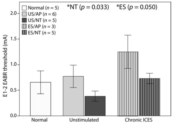
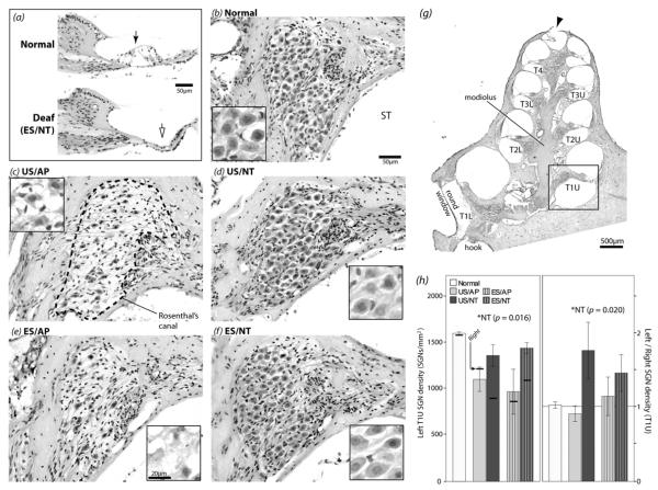
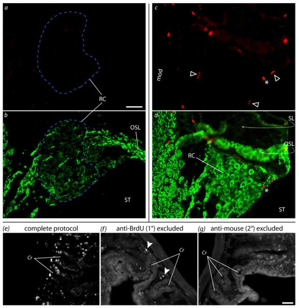
Cochlear implants electrically stimulate residual spiral ganglion neurons (SGNs) to provide auditory cues for the severe-profoundly deaf. However, SGNs gradually degenerate following cochlear hair cell loss, leaving fewer neurons available for stimulation. Providing an exogenous supply of neurotrophins (NTs) has been shown to prevent SGN degeneration, and when combined with chronic intracochlear electrical stimulation (ES) following a short period of deafness (5 days), may also promote the formation of new neurons. The present study assessed the histopathological response of guinea pig cochleae treated with NTs (brain-derived neurotrophic factor and neurotrophin-3) with and without ES over a four week period, initiated two weeks after deafening. Results were compared to both NT alone and artificial perilymph (AP) treated animals. AP/ES treated animals exhibited no evidence of SGN rescue compared with untreated deafened controls. In contrast, NT administration showed a significant SGN rescue effect in the lower and middle cochlear turns (two-way ANOVA, p < 0.05) compared with AP-treated control animals. ES in combination with NT did not enhance SGN survival compared with NT alone. SGN function was assessed by measuring electrically-evoked auditory brainstem response (EABR) thresholds. EABR thresholds following NT treatment were significantly lower than animals treated with AP (two-way ANOVA, p = 0.033). Finally, the potential for induced neurogenesis following the combined treatment was investigated using a marker of DNA synthesis. However, no evidence of neurogenesis was observed in the SGN population. The results indicate that chronic NT delivery to the cochlea may be beneficial to cochlear implant patients by increasing the number of viable SGNs and decreasing activation thresholds compared to chronic ES alone.


Figures

Figure 1
Figure 1
Auditory brainstem response traces (averaged from 200 trials) to free-field click stimuli presented to the left ear of a guinea pig (a) one week prior to deafening, and (b) two weeks after deafening. The PIII-NIII wave (heavy black) was used to determine threshold. Large threshold increases (>50dB) occurred following deafening, usually to thresholds >100dB p.e. SPL.
Figure 2
Figure 2
Micro-focus x-ray from an ES/AP animal taken 28 days after implantation showing the six electrode rings of the implant in the basal turn of the left cochlea, and the lead wires exiting the bulla. Intracochlear electrode numbering is shown. The site of neurotrophin infusion was just apical to E1. RW = round window. Scale bar = 1mm.
Figure 3
Figure 3
(a) Bipolar (E1-2) EABRs evoked by biphasic current pulses recorded at three points during the treatment period in an ES/AP animal. Threshold (indicated by the bold traces) increased over time. Grey windows indicate the wave used to determine threshold (1-2ms post-stimulus, presumably corresponding to PIII-NIII of ABR; see Fig.1). Note that EABR waveform shape and amplitude were different between days. This may be due to changes over the treatment period, but is more likely related to slight differences in recording electrode placement. Threshold should not be significantly altered by electrode placement differences alone. Stimulus artifacts have been blanked out (0-1ms). (b) Mean EABR threshold change for E1-2 from day 0 to day 28 in ES animals. An increase occurred in ES/AP animals (n = 3) whereas little change occurred in ES/NT animals (n = 5). Error bars ± 1 SEM.
Figure 4
Figure 4
(a) Mean E1-2 EABR threshold for each group at the conclusion of the treatment period. Thresholds were significantly lower in NT animals compared to AP, and thresholds were significantly higher in ES compared to US (* two-way ANOVA). Data from acutely deafened normal animals are shown for comparison but were not included in the ANOVA. Data from one ES/AP and one ES/NT animal were not included as electrode 1-2 for these two animals was not available at the time of the experiment. Error bars ± 1 SEM.
Figure 5
Figure 5
H&E stained cochlear histological samples and SGN density data for upper turn 1 (boxed area in g). (a) Representative midmodiolar sections showing an intact organ of Corti in a normal cochlea (solid arrow) compared to a degenerated organ of Corti in a chronically deaf ES/NT cochlea (open arrow). (b-f) Representative images of Rosenthal’s canal (e.g. dashed line in c) from the left (treated) cochlea for each group. SGN somata appear smaller and the packing density lower in AP cochleae compared to NT and normal controls. Higher magnification images of SGN somata from different cochleae are shown in the insets. Morphological degeneration of the somata was apparent in AP animals, unlike NT-treated cochleae. (g) Low magnification micrograph of a midmodiolar section from a normal hearing left cochlea. The upper (U) and lower (L) aspect of each turn (T) is shown (e.g. T1U = upper turn 1). The perforation created at the apex to facilitate fixative diffusion can be seen (arrowhead). (h) Left cochlea mean SGN density values (left panel) and left cochlea SGN density normalized to the contralateral control cochlea (right panel) for upper turn 1, one of the locations where significantly greater density was seen in NT animals (* two-way ANOVA). For comparison, the means for right deafened untreated control cochleae are illustrated in the left panel. The means for normal controls are shown for comparison but were not included in the ANOVAs. See Table 1 for group n values. Error bars ± 1 SEM. ST = scala tympani.
Figure 6
Figure 6
Common ground impedance means are shown for all implant electrodes over the treatment period. There was no significant treatment effect so data are shown pooled across the two ES groups (total n = 10). Measurements other than day 0 and day 28 have been pooled across 3-day bins. Impedances increased significantly over time. Error bars ± 1 SEM.
Figure 7
Figure 7
(a) Representative examples of the fibrous tissue response (f.t.) in lower turn 1 scala tympani (ST) of ES/NT and ES/AP cochleae. ES/NT cochleae appeared to have a more extensive tissue reaction in the ST than ES/AP. The * show the likely chronic locations of the implants. (b) Qualitative ranks for tissue response severity plotted against the change in impedance from post-implantation to day 28 for E5 (electrode near cochlear region examined). There was no significant relationship between impedance change and rank across all ES animals or within either ES/AP or ES/NT groups (Spearman’s rho, p > 0.05). The letters in c indicate the data points for the micrographs in a and b.
Figure 8
Figure 8
Immunodetection of BrdU in cochlear and control tissue. (a,b) Representative example showing the lack of BrdU labeling in Rosenthal’s canal (RC; blue dashed line) of an ES/NT cochlea. (a) BrdU (red) and (b) neurofilament (green) in upper turn 1. SGN somata can be seen in the neurofilament channel, but no BrdU labeling was seen in SGN nuclei. (c,d) In one ES/NT cochlea, positive BrdU labeling was seen in what appear to be Schwann cell nuclei (arrowheads) in the modiolus (mod, not shown) and RC . An intense globular object (*) that was likely artifactual – possibly non-specific secondary antibody binding as was seen in negative controls (arrowheads in f) – can also be seen. No BrdU-labeled nuclei in cochlear tissue were surrounded by a neurofilament-labeled soma. The peripheral structures (i.e. OSL and SL) in c-d can be seen to be detached from the slide and folded back (curved arrows), obscuring part of RC. This illustrates the difficulties the DNA denaturation steps can create for analysis of sectioned cochlear tissue. (e-g) Positive and negative BrdU labeling controls (gut tissue). The complete labeling protocol (e) yielded positive labeling (white) in stem cell nuclei in the intestinal crypts (Cr), whereas exclusion of primary (f) or secondary antibodies (g) did not result in labeling. The image brightness has been increased in f and g to better view the gut anatomy. OSL = osseous spiral lamina, SL = spiral limbus, ST = scala tympani. Scale bars = 50μm (same scale a-d and e-g).

Similar articles

Cited by

References

    1. Abrous DN, Koehl M, Le Moal M. Adult neurogenesis: from precursors to network and physiology. Physiol Rev. 2005;85:523–569. - PubMed
    1. Adamson CL, Reid MA, Davis RL. Opposite actions of brain-derived neurotrophic factor and neurotrophin-3 on firing features and ion channel composition of murine spiral ganglion neurons. J Neurosci. 2002;22:1385–1396. - PMC - PubMed
    1. Agterberg MJ, Versnel H, de Groot JC, Smoorenburg GF, Albers FW, Klis SF. Morphological changes in spiral ganglion cells after intracochlear application of brain-derived neurotrophic factor in deafened guinea pigs. Hear Res. 2008;244:25–34. - PubMed
    1. Agterberg MJ, Versnel H, de Groot JC, van den Broek M, Klis SF. Chronic electrical stimulation does not prevent spiral ganglion cell degeneration in deafened guinea pigs. Hear Res. 2010;269:169–179. - PubMed
    1. Agterberg MJ, Versnel H, van Dijk LM, de Groot JC, Klis SF. Enhanced Survival of Spiral Ganglion Cells After Cessation of Treatment with Brain-Derived Neurotrophic Factor in Deafened Guinea Pigs. J Assoc Res Otolaryngol. 2009;10:355–367. - PMC - PubMed

Publication types

MeSH terms