. 2011 Sep 15;51(6):1249-57.
doi: 10.1016/j.freeradbiomed.2011.06.025. Epub 2011 Jun 27.

Activation of the glutaredoxin-1 gene by nuclear factor κB enhances signaling

Affiliations

Activation of the glutaredoxin-1 gene by nuclear factor κB enhances signaling

Scott W Aesif et al. Free Radic Biol Med. .

Abstract

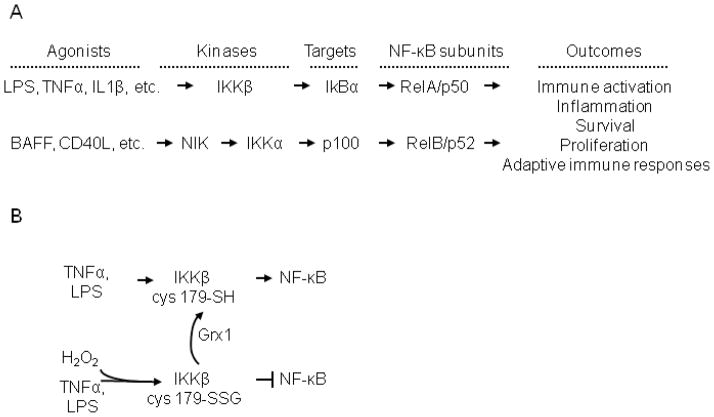
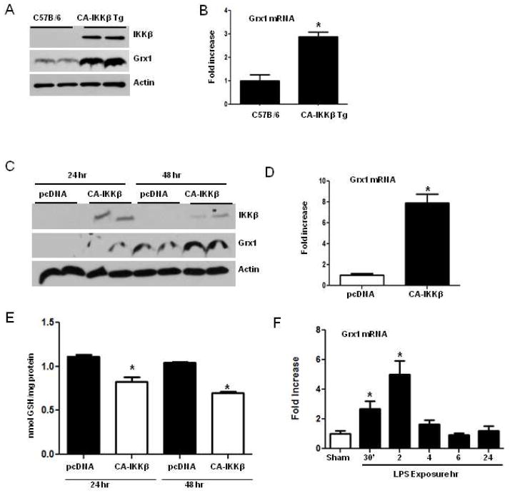
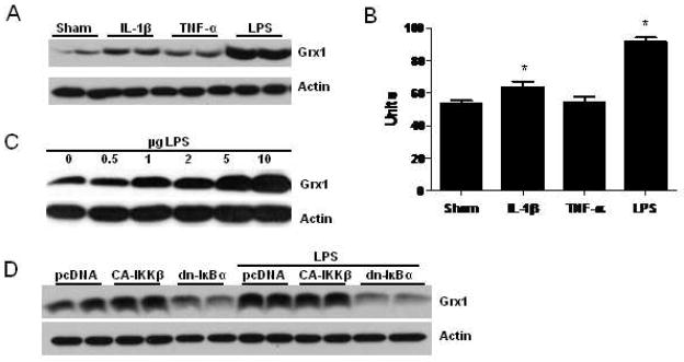
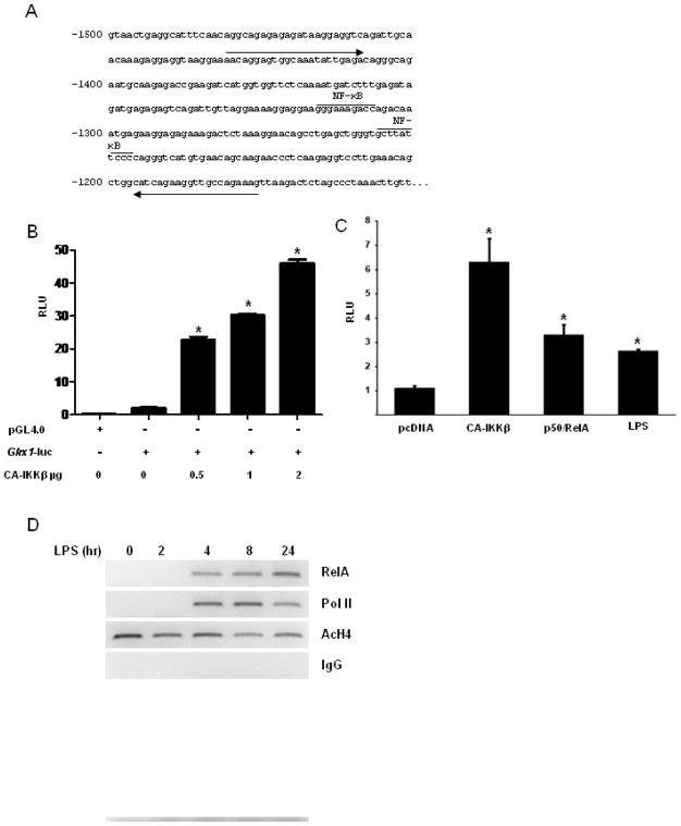
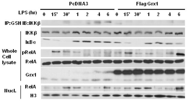
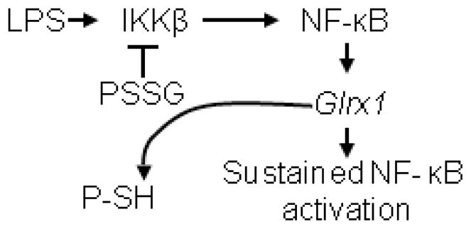
The transcription factor nuclear factor κB (NF-κB) is a critical regulator of inflammation and immunity and is negatively regulated via S-glutathionylation. The inhibitory effect of S-glutathionylation is overcome by glutaredoxin-1 (Grx1), which under physiological conditions catalyzes deglutathionylation and enhances NF-κB activation. The mechanisms whereby expression of the Glrx1 gene is regulated remain unknown. Here we examined the role of NF-κB in regulating activation of Glrx1. Transgenic mice that express a doxycycline-inducible constitutively active version of inhibitory κB kinase-β (CA-IKKβ) demonstrate elevated expression of Grx1. Transient transfection of CA-IKKβ also resulted in significant induction of Grx1. A 2-kb region of the Glrx1 promoter that contains two putative NF-κB binding sites was activated by CA-IKKβ, RelA/p50, and lipopolysaccharide (LPS). Chromatin immunoprecipitation experiments confirmed binding of RelA to the promoter of Glrx1 in response to LPS. Stimulation of C10 lung epithelial cells with LPS caused transient increases in Grx1 mRNA expression and time-dependent increases in S-glutathionylation of IKKβ. Overexpression of Grx1 decreased S-glutathionylation of IKKβ, prolonged NF-κB activation, and increased levels of proinflammatory mediators. Collectively, this study demonstrates that the Glrx1 gene is positively regulated by NF-κB and suggests a feed-forward mechanism to promote NF-κB signaling by decreasing S-glutathionylation.


Figures

Fig. 1
Fig. 1
Overview of the NF-κB activation pathways and the impact of S-glutathionylation. A) Schematic representation of activation of classical (top) and alternative (bottom) NF-κB activation pathways, and outcomes. The classical pathway is activated by diverse ligands, such as LPS, Tumor Necrosis Factor-alpha (TNFα), Interleukin 1-beta (IL-1β), among many others, which results in the activation of Inhibitory kappa B kinase beta (IKKβ) which in turn mediates degradation of IκBα, resulting in the nuclear translocation and activation of RelA/p50 NF-kB subunits. The alternative NF-κB pathway is activated by distinct subsets of ligands, such as B cell Activating Factor (BAFF), CD40 ligand (CD40L) etc. which result in NF-κB Inducing Kinase (NIK) dependent activation of I kappa B kinase alpha (IKKα) which phosphorylates p100, and resultant proteolytic processing to p52. RelB/p52 dimeric complexes then are translocated to the nucleus, to activate transcription of unique sets of genes. Note that this schematic is an oversimplification, as additional regulatory post-translational modifications, and chromatin remodeling events occur to enable transcriptional activation of genes. Cross talk between classical and alternative NF-κB pathways also occurs, and is not illustrated here. B) Impact of H2O2 of IKKβ and NF-κB signaling. Stimulation of cells with LPS or TNFα leads to activation of IKKβ, and downstream NF-κB signaling. In the presence of H2O2 (100–200 μM) or following overexpression of NOX1, IKKβ is inhibited via S-glutathionylation (-SSG) of Cys179. Overexpression of glutaredoxin-1 (Grx1) reverses S-glutathionylation of IKKβ (-SH), and permits NF-κB signaling in the presence of H2O2. This schematic is a summary of previously published data [10].
Fig. 2
Fig. 2
Increases of Grx1 expression following activation of the NF-κB pathway in lung epithelial cells. (A) Transgenic mice that express CA-IKKβ within the conducting airways, in a doxycycline inducible manner, or transgene negative littermate controls were maintained on doxycycline for 1 week. Mice were euthanized, and whole lung homogenates prepared for assessment of Grx1 expression by immunoblot analysis. IKKβ blot is shown to verify expression of trangenic IKKβ. β-actin is shown as a loading control. (B) Assessment of Grx1 mRNA content by real time PCR is lung tissues from mice expressing the CA-IKKβ transgene, compared to C57B/6 littermates. Results were normalized to the housekeeping gene, HPRT, and expressed as fold increases compared to transgene negative littermate controls that were fed doxycycline containing food. Data reflect mean +SEM of 4 mice/group. * p < 0.05 (ANOVA) compared to C57B/6 group. (C) Mouse alveolar type II cells (C10) were transfected with 1 μg or pcDNA3 or CA-IKKβ plasmids. After 24 and 48 h, whole cell lysates were evaluated for Grx1 expression by immunoblot analysis. β-actin: loading control. (D) Assessment of Grx1 mRNA expression via real time PCR in C10 cells transfected with 1 μg or pcDNA3 or CA-IKKβ plasmids. Results were normalized to the housekeeping gene, cyclophilin, and expressed as fold increases compared to pcDNA controls. * p<0.05 (Student T Test) compared to pcDNA3 controls. (E) Assessment of protein S-glutathionylation in C10 cells following expression of CA-IKKβ. 24 or 48 post transfection with PcDNA3 or CA-IKKβ, cells were lysed and proteins precipitated for assessment of PSSG. The sodium borohydride dependent release of GSH was measured. Results are normalized to cellular protein content. * p<0.05 (ANOVA) compared to pcDNA3 controls. (F) Assessment of Grx1 mRNA expression in C10 cells exposed to 1 μg/ml of LPS for the indicated times. Results were normalized to the housekeeping gene, cyclophilin, and expressed as fold increases compared to pcDNA controls. * p<0.05 (ANOVA) compared to pcDNA3 controls.
Fig. 3
Fig. 3
Expression of Grx1 in RAW264.7 macrophage like cells following stimulation with known NF-κB agonists. (A) RAW264.7 cells were stimulated with IL-1β (5ng/mL), TNF-α (10 ng/mL), or LPS (1μg/mL) for 24 h. Whole cell lysates were resolved by SDS-PAGE and immunoblotted for Grx1, and β-actin. (B) Assessment of Grx1 activity RAW264.7 cells, 24 h post stimulation with agonists, as in A. Data are expressed as mean (±SEM) units. * p < 0.05 (ANOVA) compared to sham controls. (C) Dose dependent modulation of Grx1 content in RAW264.7 cells 24 h after stimulation with LPS. RAW264.7 cells were stimulated with the indicated concentrations of LPS, and after 24 h, whole cell lysates were prepared for Western Blot analysis. (D) RAW264.7 cells were transfected with vector control (pcDNA3.0), constitutively active IKKβ (CA-IKKβ), or dominant negative IκBα (dn-IκBα). 24 h later, cells were exposed to LPS (1 μg/ml) for an additional 24 h before immunoblot analysis for Grx1. Actin is shown as a loading control.
Fig. 4
Fig. 4
Assessment of activation of the glrx1 promoter by NF-κB. (A) Schematic depiction of the glrx1 promoter highlighting two putative NF-κB1 (p50) binding sites at −1250 base pairs (bp) and −1310 bp. Arrows indicate the primer sequences used for ChIP analysis. (B) C10 cells were transfected with vector encoding β-galactosidase, empty PGL4.0 vector, or PGL4.0 vector containing the 2000 bp sequence up stream of the glrx1 gene locus (Glrx1-luc), in the presence or absence of increasing amounts of PcDNA3, or CA-IKKβ. Cells were incubated 24 h prior to luciferase activity analysis. All data are expressed as mean (±SEM) relative light units (RLU) normalized to β-galactosidase activity. * p < 0.05 (ANOVA) compared to Glrx1-luc controls. (C) Cells were transfected with Glrx1-luc and renilla luciferase (pRL-TK), and where either co-transfected with 1 μg, pcDNA3, CA-IKKβ, or 0.5 μg RelA plus 0.5 μg p50. After 24 h, pcDNA3-transfected cells were stimulated with 1 μg/ml LPS. All cells were harvested 24 h later using the dual-luciferase reporter assay system (Promega) according to manufacturer’s instructions. Data are expressed as mean (±SEM) relative light units (RLU) normalized to Renilla activity. * p < 0.05 (ANOVA) compared to PcDNA3 controls. (D) Assessment of RelA binding to the Glrx1 promoter via ChIP analysis. RAW264.7 cells were stimulated with 1 μg/ml of LPS for the indicated times. Chromatin was crosslinked, sheared, and precipitated with antibodies recognizing RelA, RNA polymerase II (Pol II), or aceylated Histone H4. Pre-immune IgG antibody was used at a control. Immmunoprecipitated DNA was subjected to PCR analysis, using primer sequences indicated in Fig. 3A.
Fig. 5
Fig. 5
Assessment of the impact of over expression of Grx1 on LPS-induced NF-κB activation and S-glutathionylation of IKKβ (IKKβ-SSG) in C10 lung epithelial cells. Top panel: S-glutathionylation of IKKβ. At the indicated times, S-glutathionylated proteins were immunoprecipitated (IP) with anti-GSH antibody, and subjected to Western Blotting to detect IKKβ. No immunoreactivity occurred in IgG control immunoprecipitations or following decomposition of protein-S-glutathionylation with DTT (data not shown). Whole cell lysates (WCL); Assessment of IKKβ content as a control in samples used for IP, and IκBα content, and phoshorylation of RelA at serine 536 (pRelA) as measures of NF-κB activation. Total RelA: loading control, Grx1: confirmation of Grx1 overexpression. Bottom panels: Assessment of nuclear content (Nucl) of RelA in response to LPS in cells transfected with PcDNA3 (left), or Grx1 (right). H3: histone H3 as a loading control.
Fig. 6
Fig. 6
Enhanced wound closure in CA-IKKβ expressing cells requires the presence of Grx1. C10 cells were transfected with control SiRNA, or Grx1 SiRNA, and 24 h thereafter transfected with PcDNA3 or CA-IKKβ. 24 h later, a scratch was made with a pipet tip, and 24 h thereafter, the % closure of the wound area quantified. Results are representative of 6 observations conducted in two separate experiments. * p < 0.05 (ANOVA) compared to the pcDNA group; ‡ p < 0.05 (ANOVA) compared to the control siRNA, CA-IKKβ-transfected group.
Fig. 7
Fig. 7
Model depicting the potential impact of Grx1 on prolonging activation of NF-κB. In response to stimulation with LPS, S-glutathionylation (PSSG) of IKKβ is important to shut down the activity of NF-κB. Activation of the Glrx1 gene via canonical NF-κB activation prevents the accumulation of IKKβ-SSG, thereby prolonging activation of the NF-κB pathway, and the production of pro-inflammatory mediators. Note that Grx1-catalyzed deglutathionylation results in the formation of protein sulfhydryl groups (P-SH). It is plausible that in addition to IKKβ, other members of the NF-κB pathway are regulated via S-glutathionylation and Grx1-catalyzed deglutationylation (not shown).

References

    1. Karin M, Greten FR. NF-kappaB: linking inflammation and immunity to cancer development and progression. Nat Rev Immunol. 2005;5:749–759. - PubMed
    1. Ghosh S, Hayden MS. New regulators of NF-kappaB in inflammation. Nat Rev Immunol. 2008;8:837–848. - PubMed
    1. Hayden MS, Ghosh S. Shared principles in NF-kappaB signaling. Cell. 2008;132:344–362. - PubMed
    1. Karin M, Lin A. NF-kappaB at the crossroads of life and death. Nat Immunol. 2002;3:221–227. - PubMed
    1. Janssen-Heininger YM, Mossman BT, Heintz NH, Forman HJ, Kalyanaraman B, Finkel T, Stamler JS, Rhee SG, van der Vliet A. Redox-based regulation of signal transduction: principles, pitfalls, and promises. Free Radic Biol Med. 2008;45:1–17. - PMC - PubMed

Publication types

MeSH terms