

Visualizing HIV-1 assembly
- PMID: 21762796
- PMCID: PMC3144478
- DOI: 10.1016/j.jmb.2011.04.062
Visualizing HIV-1 assembly
Abstract
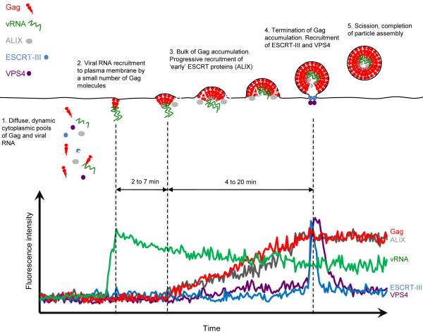
The assembly of an HIV-1 particle is a complex, multistep process involving several viral and cellular proteins, RNAs and lipids. While many macroscopic and fixed-cell microscopic techniques have provided important insights into the structure of HIV-1 particles and the mechanisms by which they assemble, analysis of individual particles and their assembly in living cells offers the potential of surmounting many of the limitations inherent in other approaches. In this review, we discuss how the recent application of live-cell microscopic imaging techniques has increased our understanding of the process of HIV-1 particle assembly. In particular, we focus on recent studies that have employed total internal reflection fluorescence microscopy and other single-virion imaging techniques in live cells. These approaches have illuminated the dynamics of Gag protein assembly, viral RNA packaging and ESCRT (endosomal sorting complex required for transport) protein recruitment at the level of individual viral particles. Overall, the particular advantages of individual particle imaging in living cells have yielded findings that would have been difficult or impossible to obtain using macroscopic or fixed-cell microscopic techniques.
Copyright © 2011 Elsevier Ltd. All rights reserved.
Figures
References
Publication types
MeSH terms
Grants and funding
LinkOut - more resources
Full Text Sources
