Review
. 2011 Jul 22;410(4):553-64.
doi: 10.1016/j.jmb.2011.04.072.

Beyond plasma membrane targeting: role of the MA domain of Gag in retroviral genome encapsidation

Affiliations
Review

Beyond plasma membrane targeting: role of the MA domain of Gag in retroviral genome encapsidation

Leslie J Parent et al. J Mol Biol. .

Abstract

The MA (matrix) domain of the retroviral Gag polyprotein plays several critical roles during virus assembly. Although best known for targeting the Gag polyprotein to the inner leaflet of the plasma membrane for virus budding, recent studies have revealed that MA also contributes to selective packaging of the genomic RNA (gRNA) into virions. In this Review, we summarize recent progress in understanding how MA participates in genome incorporation. We compare the mechanisms by which the MA domains of different retroviral Gag proteins influence gRNA packaging, highlighting variations and similarities in how MA directs the subcellular trafficking of Gag, interacts with host factors and binds to nucleic acids. A deeper understanding of how MA participates in these diverse functions at different stages in the virus assembly pathway will require more detailed information about the structure of the MA domain within the full-length Gag polyprotein. In particular, it will be necessary to understand the structural basis of the interaction of MA with gRNA, host transport factors and membrane phospholipids. A better appreciation of the multiple roles MA plays in genome packaging and Gag localization might guide the development of novel antiviral strategies in the future.


Figures

Figure. 1
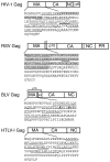
Figure. 1. Domain structure of Gag and sequences of the MA domains of RSV, HIV-1, BLV, and HTLV-1
The organization of the domains in retroviral Gag polyproteins discussed in this review (MA, matrix; CA, capsid; NC, nucleocapsid); PR (protease). The amino acid sequences of each MA domain are indicated, with the α-helices underlined and the 310 helices underlined with double lines based on coordinates defined by three-dimensional structural analyses , -. Residues in bold-face type represent residues that are required for interactions with nucleic acids. In HIV-1 MA, the gray box denotes the highly basic region (HBR) which is involved in nucleic acid binding, PIP(4,5)2 interaction, and plasma membrane targeting. For RSV MA, the gray box delineates the membrane-binding domain and the NLS in MA. The boundaries of the helices shown for HTLV-I MA are predicted from the three-dimensional structure solved for the homologous HTLV-II MA protein. Residues indicated in bold-type in the BLV MA sequence have been implicated in genome encapsidation.
Fig. 2
Fig. 2. Model illustrating the role of the Gag MA domain in regulating gRNA binding, subcellular trafficking, genome encapsidation and particle assembly
After synthesis in the cytoplasm, Gag proteins destined to bind the gRNA for packaging are transported to specific subcellular locations. The MA domain of Gag is represented by pentagons, CA by ovals, and NC by triangles. The gRNA is a wavy black line and the ψ sequence is depicted as a red cloverleaf. For RSV Gag (green), NLSs in the MA and NC domains interact with host import factors importin-11 and the importin-α/β complex to direct Gag into the nucleus where Gag interacts with the ψ sequence on the gRNA. RSV Gag:gRNA binding induces a conformation change in RSV Gag that promotes binding to CRM1:RanGTP, facilitating nuclear export. RSV Gag forms oligomers that are transported to the plasma membrane, possibly through an interaction with the phosphoinositol PIP(4,5)2 (denoted by pink ovals in the inner leaflet of the lipid bilayer). A discontinuous hexameric lattice of RSV Gag proteins bound to gRNA assembles at the membrane, encapsidating the genome into the assembling particle. For HIV (orange), Gag interacts with its gRNA at a pericentriolar location (illustrated as a yellow star) or in the cytoplasm, inducing Gag dimer formation. The model illustrates the MA and NC domains interacting with gRNA in an extended conformation (top) or in a folded conformation (bottom), with the NC domain binding to ψ and the MA domain binding to the gRNA at a different location. It is possible that the MA domain is bound to a cellular RNA rather than to the gRNA. HIV-1 Gag:gRNA oligomers form and are transported toward the periphery of the cell. Upon binding to PIP(4,5)2, the MA domain releases the gRNA and Gag adopts an extended conformation with MA facing the membrane and NC binding to the gRNA. The hexamers of Gag associate with the plasma membrane and assemble into a hexameric lattice. For BLV (blue), the site of Gag:gRNA complex formation is not known. The MA domain of BLV Gag is shown binding to the ψ sequence, although the NC domain also contacts the gRNA through nonspecific interactions. Further details regarding the mechanism of BLV Gag assembly are not well understood. For HTLV-I, the model depicts the ψ sequence on the gRNA binding to the NC domain of Gag because it is not known whether MA plays a role in genome encapsidation. It has been shown that nucleic acid binding does not influence membrane binding, and PIP(4,5)2 is not required for membrane targeting

Similar articles

Cited by

References

    1. Swanstrom R, Wills JW. Synthesis, assembly, and processing of viral proteins. In: Coffin JM, Hughes SH, Varmus HE, editors. Retroviruses. Cold Spring Harbor Laboratory Press; 1997. pp. 263–334. - PubMed
    1. Berkowitz R, Fisher J, Goff SP. RNA packaging. Curr.Top.Microbiol.Immunol. 1996;214:177–218. - PubMed
    1. Rein A. Retroviral RNA packaging: a review. Arch.Virol.Suppl. 1994;9:513–522. - PubMed
    1. Jewell NA, Mansky LM. In the beginning: genome recognition, RNA encapsidation and the initiation of complex retrovirus assembly. J Gen Virol. 2000;81(Pt 8):1889–1899. - PubMed
    1. Muriaux D, Mirro J, Harvin D, Rein A. RNA is a structural element in retrovirus particles. Proc Nat Acad Sci USA. 2001;98:5246–5251. - PMC - PubMed

Publication types

Substances

LinkOut - more resources