Review
. 2012 Mar;143(3):656-64.
doi: 10.1016/j.jtcvs.2011.04.044. Epub 2011 Jul 16.

Myocardial remodeling with aortic stenosis and after aortic valve replacement: mechanisms and future prognostic implications

Affiliations
Review

Myocardial remodeling with aortic stenosis and after aortic valve replacement: mechanisms and future prognostic implications

William M Yarbrough et al. J Thorac Cardiovasc Surg. 2012 Mar.

Abstract

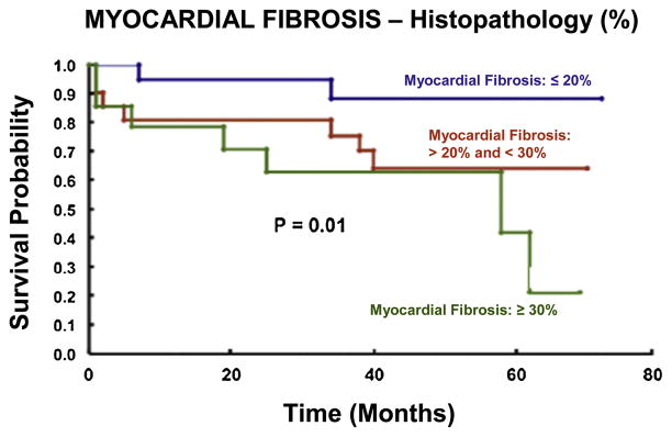
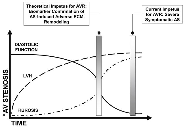
Aortic valve stenosis is a common cause of left ventricular pressure overload, a pathologic process that elicits myocyte hypertrophy and alterations in extracellular matrix composition, both of which contribute to increases in left ventricular stiffness. However, clinical and animal studies suggest that increased myocardial extracellular matrix fibrillar collagen content occurs later in the time course of left ventricular pressure overload at a time coincident with severe abnormalities in diastolic function followed by the development of symptomatic heart failure. Aortic valve replacement remains the most effective treatment for elimination of chronic pressure overload secondary to aortic stenosis but has traditionally been recommended only after the onset of clinical symptoms. Long-term follow-up of patients with symptomatic aortic stenosis after aortic valve replacement suggests that valve replacement may not result in complete reversal of the maladaptive changes that occur within the myocardial extracellular matrix secondary to the pressure overload state. To the contrary, residual left ventricular extracellular matrix abnormalities such as these are likely responsible for persistent abnormalities in diastolic function and increased morbidity and mortality after aortic valve replacement. Defining the mechanisms and pathways responsible for regulating the myocardial extracellular matrix during the natural history of aortic stenosis may provide a means by which to detect crucial structural milestones and thereby permit more precise identification of the development of maladaptive left ventricular remodeling.


Figures

Figure 1
Figure 1
Villari and associates demonstrated that regression of AS-induced cellular and extracellular abnormalities occurs in a time dependent manner post-AVR. Moreover, elimination of the pressure overload state failed to achieve normalization of myocyte diameter and interstitial fibrosis. Expressed as a percentage change from control values, AVR was associated with significant reductions in muscle fiber diameter at both early (22±8 months) and late (81±24 months) time-points compared to pre-operative values; however, muscle fiber diameter remained significantly elevated over control values late after AVR. Values for interstitial fibrosis increased early following AVR compared to pre-operative values. This increase was likely “relative” and a result of a concomitant early reduction in myocyte mass as opposed to an increase in collagen synthesis post-AVR. Values for interstitial fibrosis remained significantly elevated above those observed in the control group late following AVR. Data are presented as the mean and the standard error of the mean and were modified from Villari B, Vassalli G, Monrad ES, et al. Circulation. 91:2353–2358.1995 (*p<0.05 vs. Control, +p<0.05 vs. Pre-Op, #p<0.05 vs. Early).
Figure 2
Figure 2
Increasing degrees of myocardial fibrosis secondary to aortic valve disease (as assessed histologically) are associated with progressive reductions in survival in patients treated with AVR utilizing traditional criteria for intervention (i.e. symptom onset). Modified from Azevedo CF, Nigri M, Higuchi ML, et al. J Am Coll Cardiol. 56:278–87. 2010
Figure 3
Figure 3
The hypothetical schematic depicts deterioration in diastolic function and an increase in myocyte hypertrophy and myocardial fibrosis over time and in the setting of AS-induced progressive pressure overload. Current management paradigms advocate AVR for patients with severe AS and associated symptoms, namely, angina, syncope, and chronic heart failure. However, AVR carried out only after symptom onset likely fails to reverse the maladaptive ECM remodeling that has already occurred. Identification of a biomarker capable of heralding the transition from AS-induced adaptive to maladaptive ECM remodeling processes may therapeutic value. Specifically, identification of such a biomarker may be used to direct more timely elimination of the pressure overload state thus optimizing the chances for normalization within the ECM and improved post-operative outcomes.

Similar articles

Cited by

References

    1. Grossman W, Jones D, McLaurin LP. Wall stress and patterns of hypertrophy in the human left ventricle. J Clin Inves. 1975;53:332–41. - PMC - PubMed
    1. Badeer HS. Biological significance of cardiac hypertrophy. Am J Cardiol. 1964;14:133–138. - PubMed
    1. Alpert NR. Cardiac hypertrophy. Academic Press, Inc; New York: 1971. Preface; pp. XV–XVI.
    1. Carabello BA, Paulus WJ. Aortic Stenosis. Lancet. 2009;373:956–66. - PubMed
    1. Monrad ES, Hess OM, Murakami T, et al. Time course of regression of left ventricular hypertrophy after aortic valve replacement. Circulation. 1988;77(6):1345–55. - PubMed

Publication types

MeSH terms

Substances