Reversal of atopic dermatitis with narrow-band UVB phototherapy and biomarkers for therapeutic response
- PMID: 21762976
- PMCID: PMC3448950
- DOI: 10.1016/j.jaci.2011.05.042
Reversal of atopic dermatitis with narrow-band UVB phototherapy and biomarkers for therapeutic response
Abstract
Background: Atopic dermatitis (AD) is a common inflammatory skin disease exhibiting a predominantly T(H)2/"T22" immune activation and a defective epidermal barrier. Narrow-band UVB (NB-UVB) is considered an efficient treatment for moderate-to-severe AD. In patients with psoriasis, NB-UVB has been found to suppress T(H)1/T(H)17 polarization, with subsequent reversal of epidermal hyperplasia. The immunomodulatory effects of this treatment are largely unknown in patients with AD.
Objective: We sought to evaluate the effects of NB-UVB on immune and barrier abnormalities in patients with AD, aiming to establish reversibility of disease and biomarkers of therapeutic response.
Methods: Twelve patients with moderate-to-severe chronic AD received NB-UVB phototherapy 3 times weekly for up to 12 weeks. Lesional and nonlesional skin biopsy specimens were obtained before and after treatment and evaluated by using gene expression and immunohistochemistry studies.
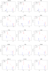
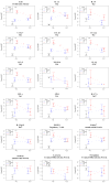
Results: All patients had at least a 50% reduction in SCORAD index scores with NB-UVB phototherapy. The T(H)2, T22, and T(H)1 immune pathways were suppressed, and measures of epidermal hyperplasia and differentiation normalized. The reversal of disease activity was associated with elimination of inflammatory leukocytes and T(H)2/T22- associated cytokines and chemokines and normalized expression of barrier proteins.
Conclusions: Our study shows that resolution of clinical disease in patients with chronic AD is accompanied by reversal of both the epidermal defects and the underlying immune activation. We have defined a set of biomarkers of disease response that associate resolved T(H)2 and T22 inflammation in patients with chronic AD with reversal of barrier pathology. By showing reversal of the AD epidermal phenotype with a broad immune-targeted therapy, our data argue against a fixed genetic phenotype.
Copyright © 2011 American Academy of Allergy, Asthma & Immunology. Published by Mosby, Inc. All rights reserved.
Figures


References
-
- Guttman-Yassky E, Lowes MA, Fuentes-Duculan J, Whynot J, Novitskaya I, Cardinale I, et al. Major differences in inflammatory dendritic cells and their products distinguish atopic dermatitis from psoriasis. J Allergy Clin Immunol. 2007;119:1210–7. - PubMed
-
- Fartasch M, Ponec M. Improved barrier structure formation in air-exposed human keratinocyte culture systems. J Invest Dermatol. 1994;102:366–74. - PubMed
-
- Palmer CN, Irvine AD, Terron-Kwiatkowski A, Zhao Y, Liao H, Lee SP, et al. Common loss-of-function variants of the epidermal barrier protein filaggrin are a major predisposing factor for atopic dermatitis. Nat Genet. 2006;38:441–6. - PubMed
Publication types
MeSH terms
Substances
Grants and funding
LinkOut - more resources
Full Text Sources
Other Literature Sources
Medical
Molecular Biology Databases

