. 2011 Sep;105(9):1226-37.
doi: 10.1016/j.jinorgbio.2011.06.003. Epub 2011 Jun 24.

Binding kinetics of calmodulin with target peptides of three nitric oxide synthase isozymes

Affiliations

Binding kinetics of calmodulin with target peptides of three nitric oxide synthase isozymes

Gang Wu et al. J Inorg Biochem. 2011 Sep.

Abstract

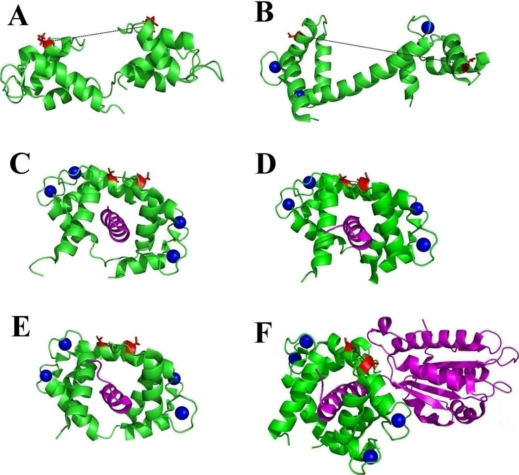
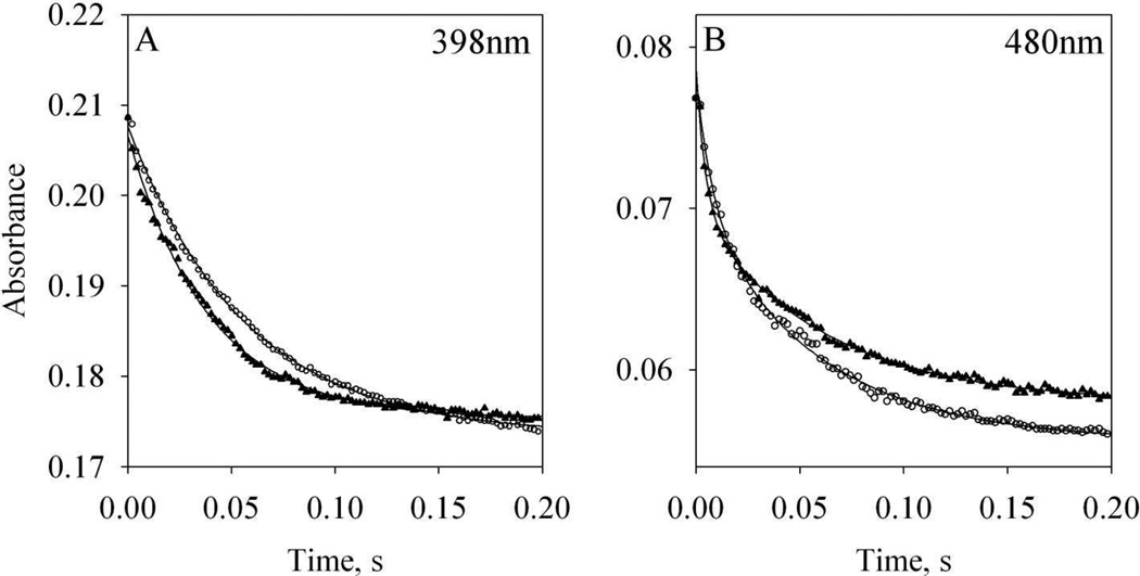
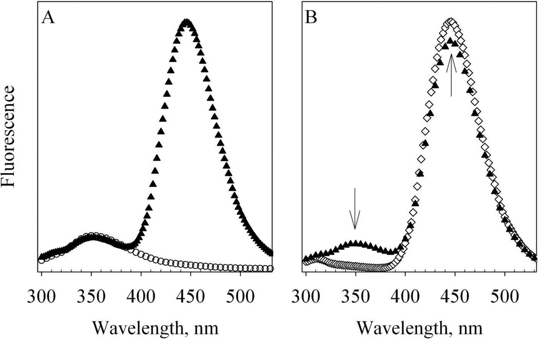
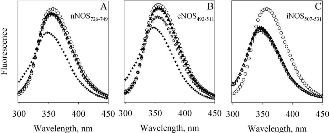
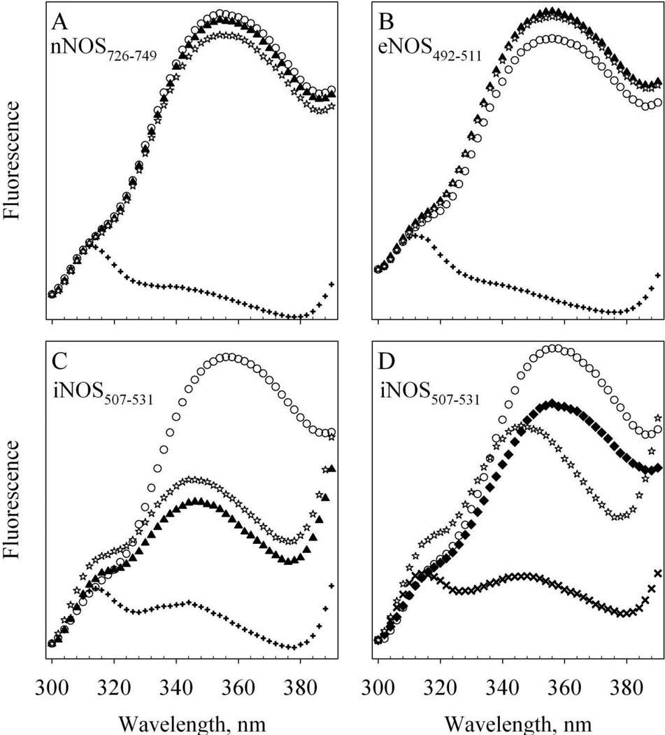
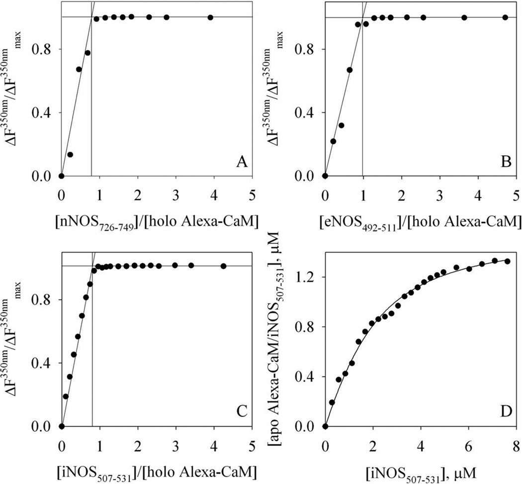
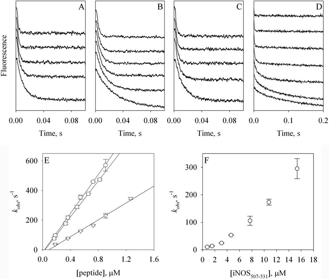
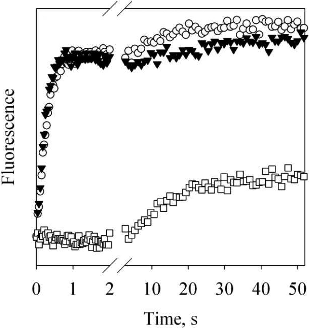
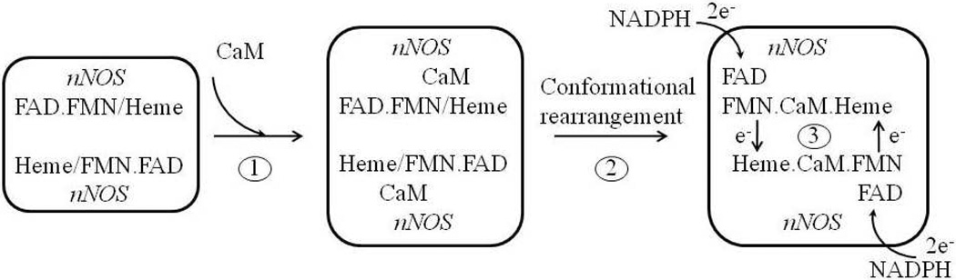
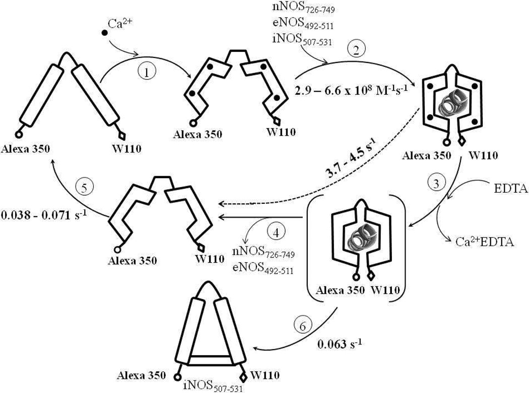
Efficient electron transfer from reductase domain to oxygenase domain in nitric oxide synthase (NOS) is dependent on the binding of calmodulin (CaM). Rate constants for the binding of CaM to NOS target peptides was only determined previously by surface plasmon resonance (SPR) (Biochemistry 35, 8742-8747, 1996) suggesting that the binding of CaM to NOSs is slow and does not support the fast electron transfer in NOSs measured in previous and this studies. To resolve this contradiction, the binding rates of holo Alexa 350 labeled T34C/T110W CaM (Alexa-CaM) to target peptides from three NOS isozymes were determined using fluorescence stopped-flow. All three target peptides exhibited fast k(on) constants at 4.5°C: 6.6×10(8)M(-1)s(-1) for nNOS(726-749), 2.9×10(8)M(-1)s(-1) for eNOS(492-511) and 6.1×10(8)M(-1)s(-1) for iNOS(507-531), 3-4 orders of magnitude faster than those determined previously by SPR. Dissociation rates of NOS target peptides from Alexa-CaM/peptide complexes were measured by Ca(2+) chelation with ETDA: 3.7s(-1) for nNOS(726-749), 4.5s(-1) for eNOS(492-511), and 0.063s(-1) for iNOS(507-531). Our data suggest that the binding of CaM to NOS is fast and kinetically competent for efficient electron transfer and is unlikely rate-limiting in NOS catalysis. Only iNOS(507-531) was able to bind apo Alexa-CaM, but in a very different conformation from its binding to holo Alexa-CaM.


Figures

Figure 1
Figure 1. Structures of CaM and its complexes with NOS targets
A: solution structure of apo CaM, 1CFC [9]; B: crystal structure of holo CaM, 1UP5 [10]; C – E, crystal structures of holo CaM complexed with NOS target peptides, C: nNOS, 2O60; D: eNOS, 1NIW [29] and E: iNOS, 3GOF. F: crystal structure of complex CaM/iNOSCaM-FMN, 3HR4 [30]. CaM molecules are shown with green ribbons and its NOS target molecules are shown with magenta ribbons. Calcium ions are represented with blue spheres. Residues T34 (left in each structure) and T110 (right in each structure) are represented with red sticks. The distances between Cα atoms of T34 and T110 are measured from the structures and indicated in black dash lines: 27.0 Å in apo CaM, 51.5 Å in holo CaM. This distance is 12.4, 13.0 and 13.3 Å in structures 2O60, 1NIW and 3GOF, respectively. In structure 3HR4, this distance is 13.6 Å.
Figure 2
Figure 2. Stopped-flow kinetics of heme and flavin reduction in wt nNOS by NADPH
Open circles: kinetics of 2µM wt nNOS replenished with 100 µM L-arginine and 5 µM BH4 reacted with a mixture of 50 µM NADPH and 2 µM holo wt CaM at room temperature. Solid triangles: 2µM wt nNOS replenished with 100 µM L-arginine and 5 µM BH4 was incubated with 2 µM holo wt CaM and then reacted with 50 µM NADPH. Each kinetic profile was an average of three stopped-flow traces. A: reduction of heme followed at 398 nm; B: reduction of flavin followed at 480 nm. Lines represent the fittings to Eqs. (1) and (2).
Figure 3
Figure 3. Fluorescence spectra of T34C/T110W CaM and Alexa-CaM
A. 1.3µM apo T34C/T110W CaM (open circles) and 1.3µM apo Alexa-CaM (solid triangles) at room temperature. Buffer: 50 mM HEPES, pH 8.0 with 100µM EDTA. B. 1.3µM holo Alexa-CaM in 50 mM HEPES, pH 8.0 with 100µM EDTA (solid triangles) and mixed with 5.8 µM eNOS492–511 and 300 µM CaCl2 (open diamonds) at room temperature. Each spectrum is the average of two scans and the fluorescence intensities are in arbitrary units. The arrows stand for the changes of fluorescence.
Figure 4
Figure 4. Fluorescence of T34C/T110W CaM and its complexes with NOS target peptides
Each spectrum is the average of two scans obtained at room temperature and the fluorescence intensities are in arbitrary units. Buffer: 50 mM HEPES, pH 7.8 with 100 mM NaCl and 50 µM EDTA. 8.7µM Apo T34C/T110W CaM (open circles) was first mixed with (A) 39.3 µM nNOS726–749 (B) 35.3 µM eNOS492–511 or (C) 31 µM iNOS507–531 (solid triangles); then mixed with 450 µM CaCl2 (plus) and finally mixed with 4 mM EDTA (stars).
Figure 5
Figure 5. Fluorescence of Alexa-CaM and its complexes with NOS target peptides
The change of FW110 between 300 – 390 nm at room temperature is shown. Each spectrum is the average of two scans and the fluorescence intensities are in arbitrary units. Buffer: 50 mM HEPES, pH 7.8 with 100 mM NaCl and 100 µM EDTA. A – C: apo 53 nM Alexa-CaM (open circles) was first mixed with (A) 3.4 µM nNOS726–749 (B) 3.4 µM eNOS492–511 or (C) 3.1 µM iNOS507–531 (solid triangles); then mixed with 300 µM CaCl2 (plus) and finally mixed with 2 mM EDTA (stars). D: apo 53 nM Alexa-CaM (open circles) was first mixed with 300 µM CaCl2 (solid diamonds); then mixed with 3.1 µM iNOS507–531 (cross) and finally with 2 mM EDTA (stars).
Figure 6
Figure 6. Titration of Alexa-CaM with NOS target peptides
A – C: titration of holo Alexa-CaM with NOS target peptides at room temperature. The percentage of FW110 (in arbitrary units) change at 350 nm is plotted versus the ratio, [NOS peptide] : [holo Alexa-CaM]. A: nNOS726–749; B: eNOS492–511; C: iNOS507–531. The lines are the visual guides for determining the breaking points of the titrations. The concentrations of holo Alexa-CaM were 1.3 µM for A and B, and 0.9 µM for C. Buffer: 50 mM HEPES, pH 8.0 with 100 mM NaCl and 200 µM CaCl2. D: titration of 1.3 µM apo Alexa-CaM with iNOS507–531 at room temperature. Buffer: 50 mM HEPES, pH 8.0 with 100 mM NaCl and 2 mM EDTA. The concentration of complex apo Alexa-CaM/iNOS507–531 is plotted versus [iNOS507–531] and the line represents the fit to Equation (4).
Figure 7
Figure 7. Kinetics of the binding of Alexa-CaM to NOS target peptides
A – D: stopped-flow time-dependent FW110 change at 4.5 °C during the association of holo Alexa-CaM with nNOS726–749 (A), eNOS492–511 (B) or iNOS507–531 (C), or the association of apo Alexa-CaM with iNOS507–531 (D). A – C: [holo Alexa-CaM] = 44 nM and [peptide] = 0.18 to 1.3 µM (bottom to top). D: [apo Alexa-CaM] = 44 nM and [peptide] = 0.8 to 15.3 µM (bottom to top). Traces are vertically offset for clarity. Buffer: 50 mM HEPES, pH 8.0 with 100 mM NaCl and 200 µM CaCl2 (A –C) or 50 mM HEPES, pH 8.0 with 100 mM NaCl and 500 µM EDTA (D). Each trace was an average of 8 – 14 stopped-flow shots. E: dependence of the rate constants kobs of holo Alexa-CaM on the concentrations of the peptides: nNOS726–749 (circles), eNOS492–511 (triangles) or iNOS507–531 (squares). F: dependence of the rate constants kobs of apo Alexa-CaM on the concentrations of iNOS507–531. The error bars are from two individual measurements.
Figure 8
Figure 8. Dissociation of Alexa-CaM from NOS target peptides
Time-dependent changes of FW110 upon mixing holo Alexa-CaM/NOS target peptide complexes with 2 mM EDTA at 4.5 °C. Starting complexes: 44 nM holo Alexa-CaM with 2.5 µM NOS peptide: nNOS726–749 (open circles), eNOS492–511 (solid triangles) or iNOS507–531 (open squares). Each trace was an average of 8 – 14 stopped-flow shots. Data between 2 s and 2.5 s were omitted for clarity.
Scheme 1
Scheme 1. CaM binding facilitates electron transfer in nNOS
Each monomer of nNOS homodimer binds one CaM in the presence of Ca2+ in Step 1. Conformational rearrangement presumably happens subsequently (Step 2) to position FMN properly for fast electron transfer from FMN to heme (Step 3).
Scheme 2
Scheme 2. Kinetic events during the binding of Alexa-CaM to NOS target peptides
Values of all rate constants were determined in this study. The species in parenthesis is unresolved intermediate for Steps 3 and 4. The dashed line with rate 3.7 – 4.5 s−1 represents the composite step including Steps 3 and 4 for either nNOS726–749 or eNOS492–511 (see Discussion). The rate of Ca2+ binding to apo Alexa-CaM (Step 1) was not measured in this study but assumed to be as fast as that determined for other CaM species [57]. Holo Alexa-CaM is assumed to bind NOS peptides in classic conformation (Step 2). Distances between Alexa 350 and W110 are purely illustrative and by no means to indicate real scales. The conformation of the apo Alexa-CaM/iNOS507–531 complex is only descriptive to demonstrate that Alexa 350 and W110 are not as separated as in free apo Alexa-CaM.

References

    1. Alderton WK, Cooper CE, Knowles RG. Biochem J. 2001;357:593–615. - PMC - PubMed
    1. Daff S. Nitric Oxide. 2010;23:1–11. - PubMed
    1. Stuehr DJ, Tejero J, Haque MM. FEBS J. 2009;276:3959–3974. - PMC - PubMed
    1. Panda K, Ghosh S, Stuehr DJ. J. Biol. Chem. 2001;276:23349–23356. - PubMed
    1. Su Z, Blazing MA, Fan D, George SE. J. Biol. Chem. 1995;270:29117–29122. - PubMed

Publication types

MeSH terms

LinkOut - more resources