. 2011 Oct;141(4):1451-62, 1462.e1-6.
doi: 10.1053/j.gastro.2011.07.003. Epub 2011 Jul 18.

Duct cells contribute to regeneration of endocrine and acinar cells following pancreatic damage in adult mice

Affiliations

Duct cells contribute to regeneration of endocrine and acinar cells following pancreatic damage in adult mice

Angela Criscimanna et al. Gastroenterology. 2011 Oct.

Abstract

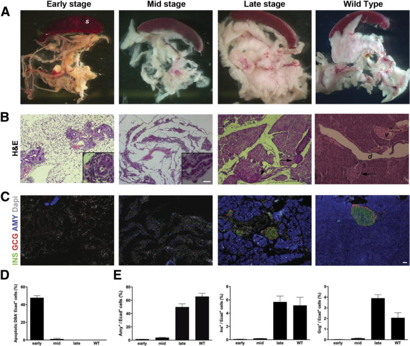
Background & aims: There have been conflicting results on a cell of origin in pancreatic regeneration. These discrepancies predominantly stem from lack of specific markers for the pancreatic precursors/stem cells, as well as differences in the targeted cells and severity of tissue injury in the experimental models so far proposed. We attempted to create a model that used diphtheria toxin receptor (DTR) to ablate specific cell populations, control the extent of injury, and avoid induction of the inflammatory response.

Methods: To target specific types of pancreatic cells, we crossed R26DTR or R26DTR/lacZ mice with transgenic mice that express the Cre recombinase in the pancreas, under control of the Pdx1 (global pancreatic) or elastase (acinar-specific) promoters.

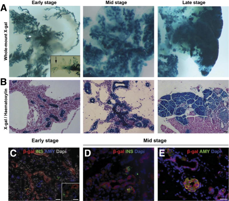
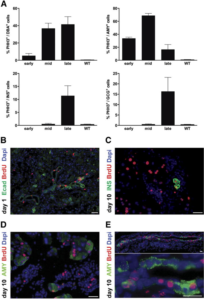
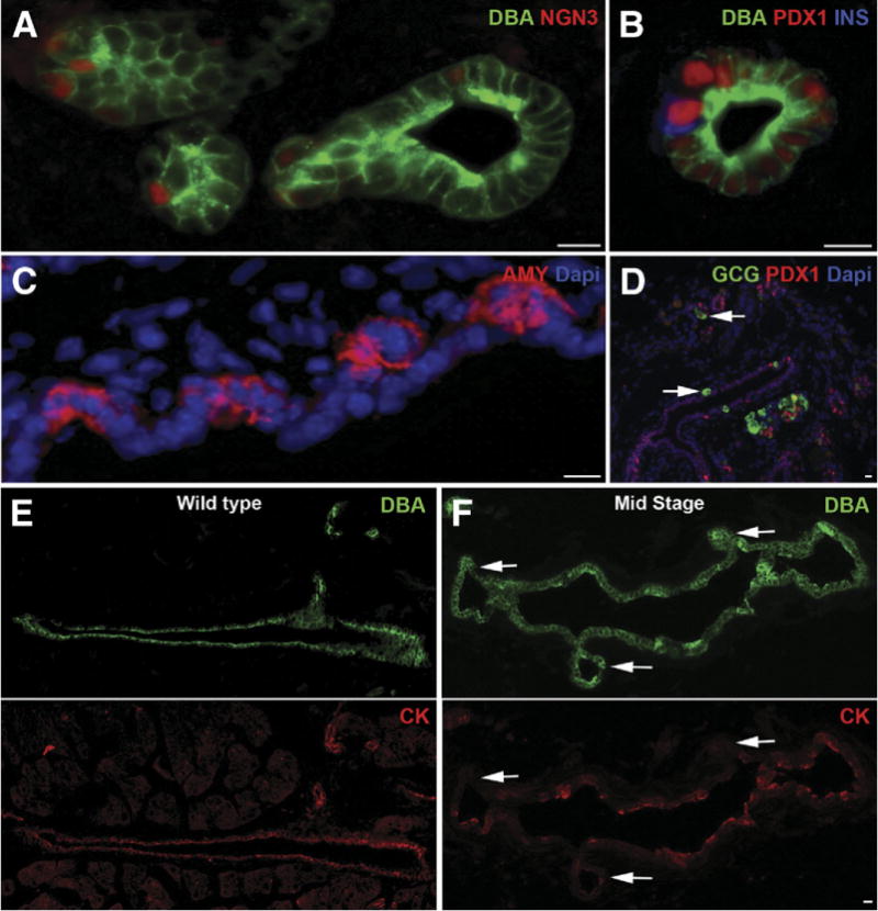
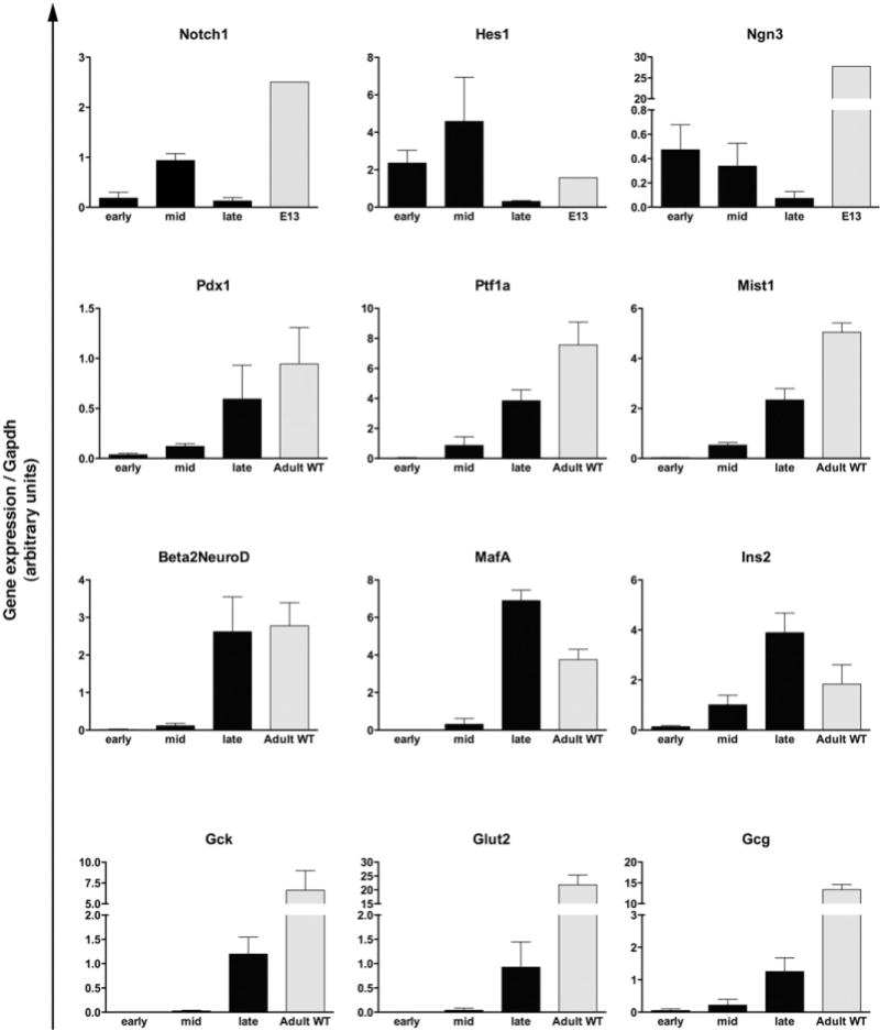
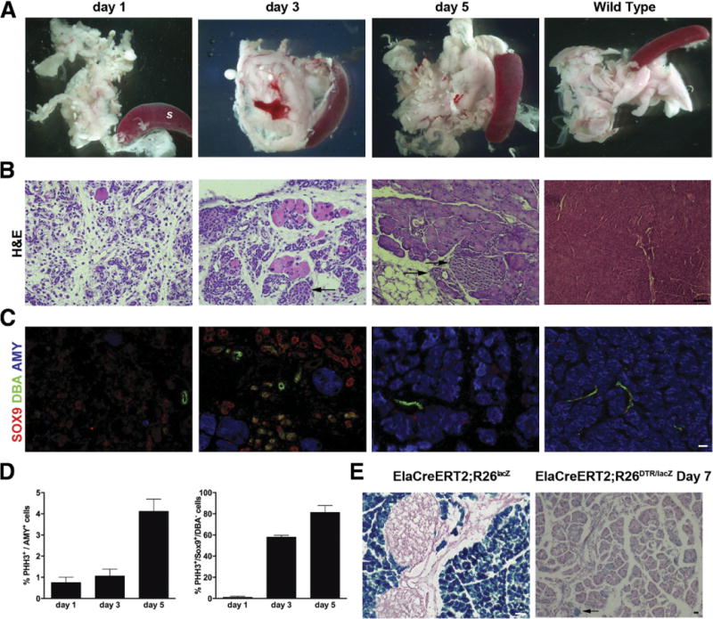
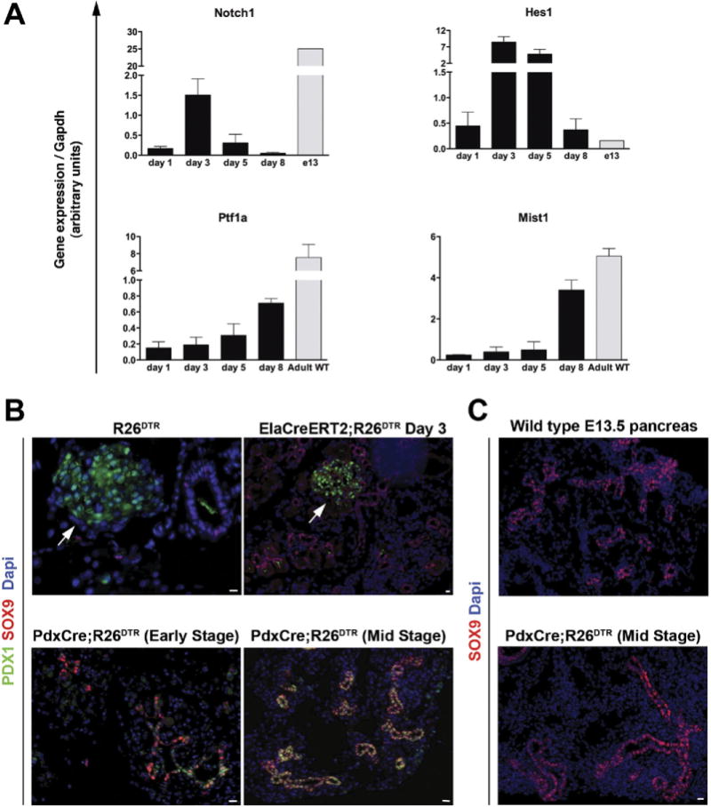
Results: Exposure of PdxCre;R26DTR mice to diphtheria toxin resulted in extensive ablation of acinar and endocrine tissues but not ductal cells. Surviving cells within the ductal compartment contributed to regeneration of endocrine and acinar cells via recapitulation of the embryonic pancreatic developmental program. However, following selective ablation of acinar tissue in ElaCreERT2;R26DTR mice, regeneration likely occurred by reprogramming of ductal cells to acinar lineage.

Conclusions: In the pancreas of adult mice, epithelial cells within the ductal compartment contribute to regeneration of endocrine and acinar cells. The severity of injury determines the regenerative mechanisms and cell types that contribute to this process.


Conflict of interest statement

Conflicts of interest

The authors disclose no conflicts.

Figures

Figure 1
Figure 1
Loss and regeneration of pancreatic mass in DT-treated PdxCre;R26DTR mice. After DT treatment, mice were killed at early, mid, or late stage. (A) Macroscopic appearance shows progressive rescue of pancreatic mass during regeneration. (B) Representative H&E staining of pancreata at the same time points. Notably, upon DT treatment, all epithelial cells were killed but ducts (insets). Arrows show islets. (C) Immunostaining for amylase, insulin, and glucagon confirmed the initial loss and subsequent regeneration of both exocrine and endocrine tissues. Wild type (WT) is an age-matched adult control pancreas. (D) Quantification of apoptotic cells (percent) among the nonductal epithelial (DBA/E+) population shows the massive cell loss after DT treatment. (E) Quantification (percent) of amylase+, insulin+, and glucagon+ cells normalized by number of E-cadherin+ cells. S, spleen; d, duct; v, vessel. Scale bars = 20 μm.
Figure 2
Figure 2
Lineage tracing analyses in the regenerating PdxCre;R26DTR/lacZ model. (A) Whole-mount X-gal staining of early-, mid-, and late-stage regenerating pancreata. Shortly after DT treatment, the only X-gal+ structures detectable were the surviving ducts, whereas mid- and late-stage staining revealed extensive contribution of X-gal+ cells to parenchymal restoration. (Inset) X-gal segment within the ductal network (arrow). (B) X-gal stained pancreata, counterstained with hematoxylin. Dashed line highlights an islet. (C) β-gal/insulin/amylase immunostaining of PdxCre;R26DTR/lacZ pancreas early after injury showed no β-gal expression in insulin+ or amylase+ cells. Inset shows higher magnification. (D) β-gal/insulin and (E) β-gal/amylase staining of mid stage PdxCre;R26DTR/lacZ pancreata showed double+ cells. Scale bars = 20 μm.
Figure 3
Figure 3
Cells within the ductal compartment are highly proliferative in regenerating PdxCre;R26DTRpancreata. (A) Quantification (percent) of proliferating DBA+, amylase+, or insulin+ cells. PHH3, phospho-histone H3. Wild type (WT) is an age-matched adult control pancreas. (B) Pulse-chase BrdU experiments. On day 1 (pulse), the duct-like structures were the main epithelial cells incorporating BrdU. On day 10 (chase), presence of (C) insulin+/BrdU+ cells and (D) amylase+/BrdU+ cells suggested that these cells or their precursors must have been proliferating mmediately after injury. (E) Several amylase+/BrdU+ cells were found within both the small and large duct-like structures, further supporting the hypothesis that the main source of regenerating acinar cells resides in the ducts rather than in proliferating residual Cre acinar cells. Scale bars = 20 μm.
Figure 4
Figure 4
Recapitulation of pancreatic developmental program in regenerating PdxCre;R26DTR pancreata. During mid stage, ductal structures showed presence of endocrine progenitor marker (A) Ngn3+, (B) PDX1+/insulin+, (C) amylase+ (AMY)+, or (D) glucagon+ (GCG) cells. A subpopulation of PDX1+/glucagon+ cells was also detected during mid stage (arrows). (E) Duct cells in wild-type pancreas were all DBA+/cytokeratin+ (CK). (F) In the regenerating pancreas, a significant number of DBA+ cells had lost cytokeratin expression (arrows). Wild type is an age-matched adult control pancreas. Scale bars = 20 μm.
Figure 5
Figure 5
Expression (qRT-PCR) of embryonic and adult pancreatic markers in the regenerating PdxCre;R26DTR pancreata. Bars represent gene expression (mean ± SE) of mice killed at early, mid, or late stage (n = 5 for each time point). Wild type (WT) is age-matched adult control pancreata (n = 5); E13 is pooled pancreata from WT litters (n = 8 embryos).
Figure 6
Figure 6
Acinar-specific cell ablation and regeneration in DT-treated ElaCreERT2;R26DTR pancreata. (A) Massive acinar cell loss was observed on day 1 after DT treatment, peaked on day 3, and was nearly complete on day 5. S, spleen. (B) Representative H&E staining of pancreata at the same time points. Arrows highlight islets. (C) Immunostainingfor DBA/SOX9/amylase in the regenerating ElaCreERT2;R26DTR pancreas showed several SOX9+/DBA duct-like structures during peak of regeneration. By contrast, ducts in wild type are DBA+/SOX9+. (D) X-gal staining showed minimal contribution of preexisting acinar cells to acinar regeneration. (E) Quantification (percent) of proliferating amylase+ and SOX9+/DBA cells. Arrows highlight X-gal+ acinar cells. Scale bars = 20 μm.
Figure 7
Figure 7
(A) Expression (qRT-PCR) of embryonic and adult exocrine markers in the regenerating ElaCreERT2;R26DTR pancreata. Bars represent gene expression (mean ± SE) of mice killed at different time points (n = 5 for each group). Wild type (WT) is age-matched adult control pancreata (n = 5); E13 is pooled pancreata from WT litters (n = 8 embryos). (B and C) The regeneration mechanism is dictated by the severity of injury. (B) Immunostaining analysis of SOX9 and PDX1 on R26DTR control pancreas, day 3 ElaCreERT2;R26DTR, and early-stage or mid-stage regenerating PdxCre;R26DTR pancreata showed coexpression of these markers only in DT-treated PdxCre;R26DTR pancreas. Arrows highlight islets. (C) SOX9 expression in the regenerating PdxCre;R26DTR pancreas displayed striking similarities to an E13.5 embryonic pancreas. Scale bars = 20 μm.

References

    1. Puri S, Hebrok M. Cellular plasticity within the pancreas—lessons learned from development. Dev Cell. 2010;18:342–356. - PMC - PubMed
    1. Bonner-Weir S, Li WC, Ouziel-Yahalom L, et al. Beta-cell growth and regeneration: replication is only part of the story. Diabetes. 2010;59:2340–2348. - PMC - PubMed
    1. Dor Y, Brown J, Martinez OI, et al. Adult pancreatic beta-cells are formed by self-duplication rather than stem-cell differentiation. Nature. 2004;429:41–46. - PubMed
    1. Teta M, Rankin MM, Long SY, et al. Growth and regeneration of adult beta cells does not involve specialized progenitors. Dev Cell. 2007;12:817–826. - PubMed
    1. Brennand K, Huangfu D, Melton D. All beta cells contribute equally to islet growth and maintenance. PLoS Biol. 2007;5:e163. - PMC - PubMed

Publication types

MeSH terms