. 2011 Sep;179(3):1287-300.
doi: 10.1016/j.ajpath.2011.05.032. Epub 2011 Jul 16.

Regression of allograft airway fibrosis: the role of MMP-dependent tissue remodeling in obliterative bronchiolitis after lung transplantation

Affiliations

Regression of allograft airway fibrosis: the role of MMP-dependent tissue remodeling in obliterative bronchiolitis after lung transplantation

Masaaki Sato et al. Am J Pathol. 2011 Sep.

Abstract

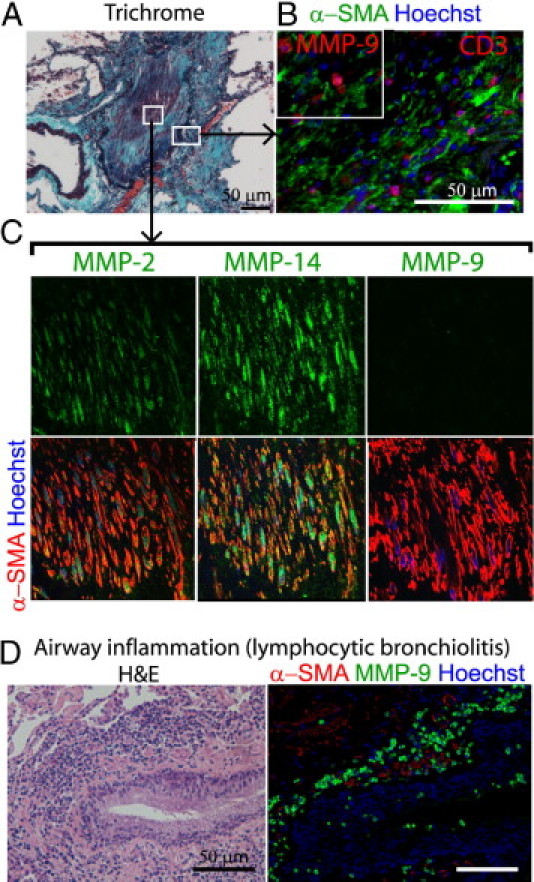
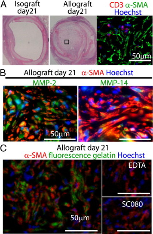
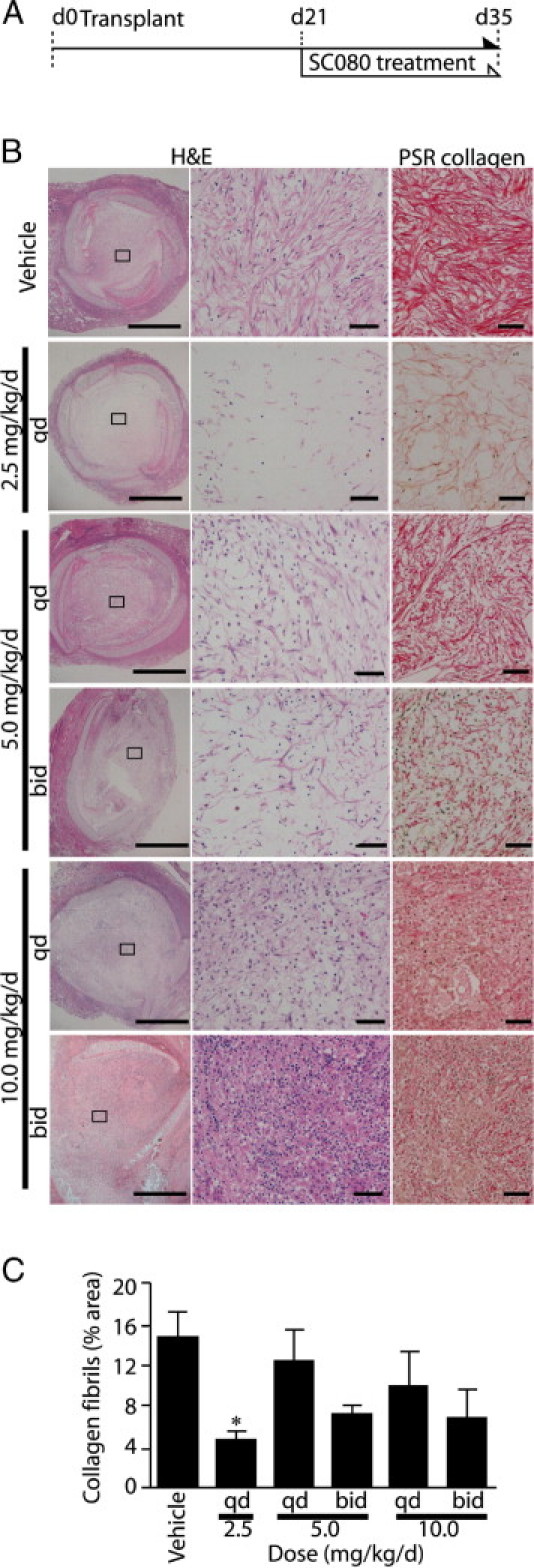
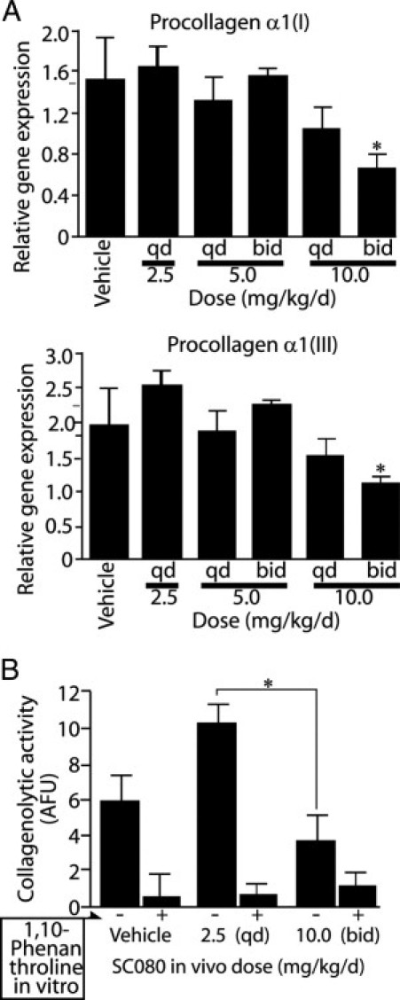
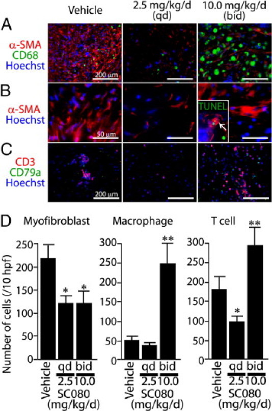
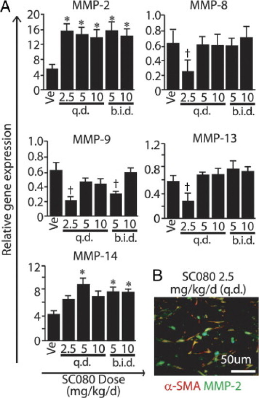
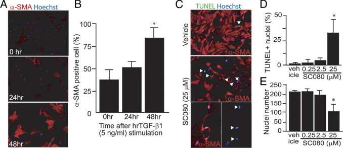
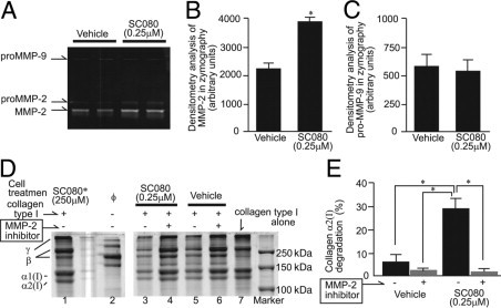
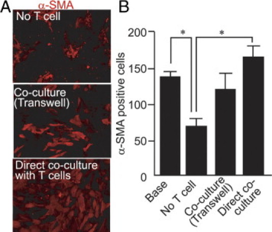
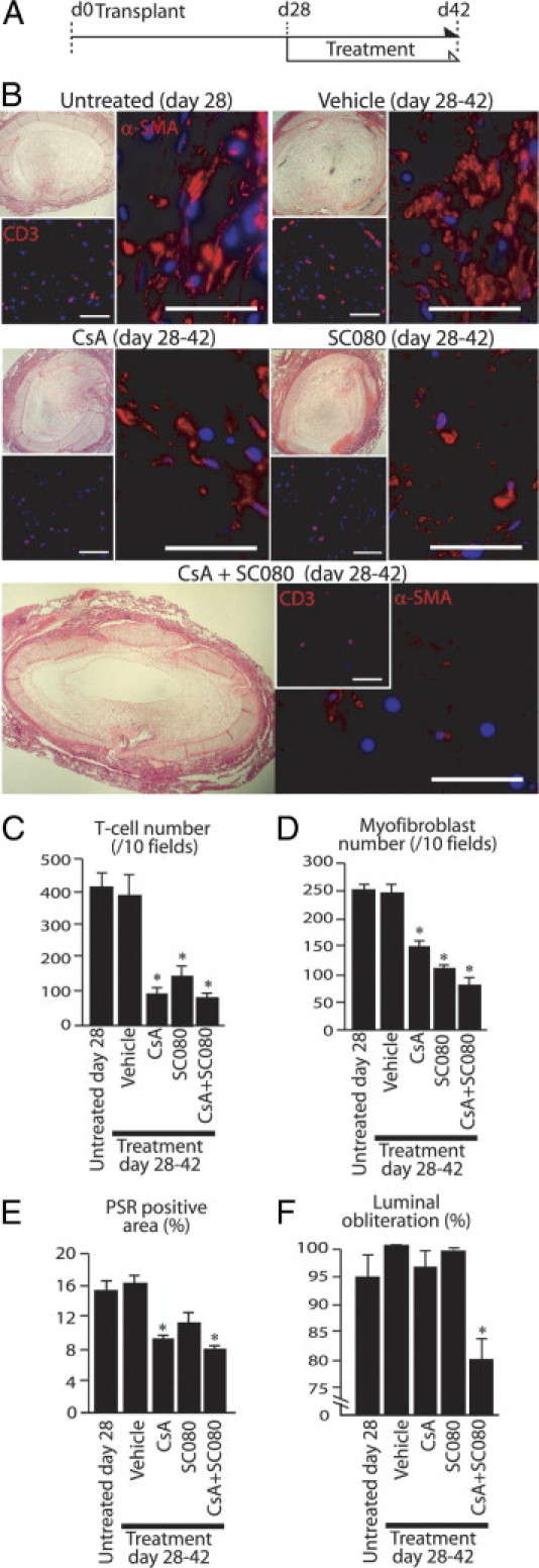
Obliterative bronchiolitis after lung transplantation is a chronic inflammatory and fibrotic condition of small airways. The fibrosis associated with obliterative bronchiolitis might be reversible. Matrix metalloproteinases (MMPs) participate in inflammation and tissue remodeling. MMP-2 localized to myofibroblasts in post-transplant human obliterative bronchiolitis lesions and to allograft fibrosis in a rat intrapulmonary tracheal transplant model. Small numbers of infiltrating T cells were also observed within the fibrosis. To modulate inflammation and tissue remodeling, the broad-spectrum MMP inhibitor SC080 was administered after the allograft was obliterated, starting at post-transplant day 21. The allograft lumen remained obliterated after treatment. Only low-dose (2.5 mg/kg per day) SC080 significantly reduced collagen deposition, reduced the number of myofibroblasts and the infiltration of T cells in association with increased collagenolytic activity, increased MMP-2 gene expression, and decreased MMP-8, MMP-9, and MMP-13 gene expression. In in vitro experiments using cultured myofibroblasts, a relatively low concentration of SC080 increased MMP-2 activity and degradation of type I collagen. Moreover, coculture with T cells facilitated persistence of myofibroblasts, suggesting a role for T-cell infiltration in myofibroblast persistence in fibrosis. By combining low-dose SC080 with cyclosporine in vivo at post-transplant day 28, partial reversal of obliterative fibrosis was observed at day 42. Thus, modulating MMP activity might reverse established allograft airway fibrosis by regulating inflammation and tissue remodeling.


Figures

Figure 1
Figure 1
Localization of MMP-2 and MMP-14 in active fibrotic human OB lesions after lung transplantation. A: Masson trichrome staining demonstrates collagen deposition in the lumen of a small airway affected by active OB. Boxed areas correspond to images at higher magnification in B and C. B: Immunofluorescence labeling for CD3 (T cells) and α-SMA (myofibroblasts) demonstrates a number of myofibroblasts and a relatively small number of infiltrating T cells. Inset: Immunofluorescence labeling for MMP-9 and α-SMA, demonstrating expression of MMP-9 by infiltrating cells among myofibroblasts. Same original magnification as the main image. C: Immunofluorescence labeling for MMP-2, −14, or −9 and α-SMA for myofibroblasts demonstrates a number of myofibroblasts in the lumen positive for MMP-2 and MMP-14 but not MMP-9. The orange color of myofibroblasts indicates colocalization of α-SMA with MMP-2 or MMP-14. Original magnification, 600×. D: A lesion of lymphocytic bronchiolitis, showing inflammatory infiltration around a bronchiole under H&E staining (left) and inflammatory cells positive for MMP-9 around the airway (right). Note that smooth muscle layers positive for α-SMA (red) around the airway are negative for MMP-9 (green). Scale bars = 50 μm.
Figure 2
Figure 2
MMP-2, MMP-14, and gelatinolytic activity in allograft airway fibrosis in an intrapulmonary tracheal transplant model. A: An isograft and an allograft at day 21 after intrapulmonary tracheal transplantation. Under H&E staining, isografts showed complete recovery of the epithelium with an open lumen (left), but the allografts showed complete luminal obliteration with fibrous tissue (middle). Immunofluorescence labeling for CD3 and α-SMA demonstrates a number of myofibroblasts and a relatively small number of infiltrating T cells (right). B: Localization of MMP-2 and MMP-14 to α-SMA+ myofibroblasts in allograft airway fibrosis at day 21. Graft-infiltrating immune cells are also positively stained for these MMPs. C: Fluorescence in situ zymography for an established allograft airway fibrosis at day 21. In situ gelatin zymography using fluorescence-quenched DQ-gelatin in combination with immunofluorescence labeling for α-SMA in a frozen tissue section demonstrates gelatinolytic activity in allograft airway fibrosis. Note the pericellular green fluorescence indicating active matrix degradation at the periphery of the cells. Negative controls including EDTA, which chelates Zn2+ necessary for MMP activity, or direct inactivation of MMPs with SC080 in vitro demonstrate that the gelatinolytic activity is mediated by MMPs. Scale bars = 50 μm.
Figure 3
Figure 3
The lowest dose of SC080 most effectively reduces extracellular matrix and myofibroblasts in the lumen. A: Five different regimens of SC080 (2.5 to 10 mg/kg day, administered q.d. or b.i.d.) were tested on allograft recipient animals from day 21 to day 35 after transplantation. B: Allograft tracheae were histologically examined at day 35 using H&E and PSR collagen staining. Boxed areas in the left column correspond to images at higher magnification in the middle and right columns. Both treated and control allografts showed luminal obliteration. Animals treated with a low dose of SC080 (2.5 mg/kg per day, q.d.) showed the lowest amount of ECM and the least cellularity. Animals treated with intensive SC080 (10 mg/kg per day, b.i.d.) showed increased mononuclear cell infiltration. Treatment with intermediate doses resulted in moderate changes in cellularity and ECM deposition. Scale bars: 500 μm (left column); 50 μm (middle and right columns). C: Morphometric quantification of PSR staining under polarized light demonstrated significantly reduced total collagen in low-dose SC080 treatment. *P < 0.05 versus vehicle control, Tukey's test, n = 5 for each group.
Figure 4
Figure 4
Low-dose SC080 treatment increases MMP-dependent collagenolytic activity without changing procollagen gene expression. A: Quantitative real-time RT-PCR analysis for gene expression of procollagen α1(I) and α1(III), the precursors for type I and type III collagen, respectively. Both genes showed dose-dependent decreases in expression. P < 0.01, regression analysis. The high-dose treatment showed a significant decrease in procollagen α1(I) and α1(III) gene expression. *P < 0.05, analysis of variance followed by a Tukey post hoc analysis. B: Collagenolytic activity in tissue homogenates of allograft tracheae for control, low-dose SC080 treatment (2.5 mg/kg per day, q.d.) and intensive SC080 treatment (10 mg/kg per day, b.i.d.). Collagenolytic activity was significantly higher in the low-dose SC080 group than in intensive SC080 group. *P < 0.05, n = 5 for each group.
Figure 5
Figure 5
SC080 treatment modifies cellular components of allograft airway fibrosis. A: Immunofluorescence labeling for myofibroblasts (α-SMA) and macrophages (CD68). B: Immunofluorescence labeling for myofibroblasts (α-SMA). Note the irregular-shaped myofibroblasts with scattered distribution in the low-dose group and the large, well-stretched myofibroblasts in the intensive treatment group compared with those in controls. Inset: Immunofluorescence with TUNEL; the arrow indicates a TUNEL-positive myofibroblast. Same original magnification as the main image. C: Immunofluorescence labeling for T cells (CD3) and B cells (CD79a). The groups of low-dose (2.5 mg/kg per day, q.d.) and intensive (10 mg/kg per day, b.i.d.) SC080 treatment and vehicle control were examined. D: Semiquantitative analysis of the numbers of myofibroblasts, macrophages, and T cells in the fibrotic area of the graft lumen. The number of myofibroblasts was smaller in the low-dose and intensive SC080 treatments (*P < 0.05). T-cell numbers in the low-dose group were significantly smaller than in the control group (*P < 0.05), whereas the numbers of macrophages and T cells were significantly larger in the intensive SC080 treatment group than in the control group (**P < 0.05). n = 5 for each group. Scale bars: 200 μm (A and C); 50 μm (B).
Figure 6
Figure 6
Increased MMP-2 gene expression and reduced inflammation-related MMPs in low-dose SC080 treatment. A: Gene expression analysis of MMPs by real-time RT-PCR. Gene expression of MMP-2 in the low-dose SC080 group was significantly higher than that in controls (*P < 0.05) and gene expression of MMP-14 was not significantly different, whereas gene expression of MMP-8, −9, and −13 in the low-dose SC080 group was significantly lower than in controls (P < 0.05). B: Double immunofluorescence labeling for α-SMA and MMP-2. MMP-2 was localized mainly to myofibroblasts positive for α-SMA in the fibrous tissue of allograft airway lumen in the low-dose SC080 group and in vehicle controls. Scale bar = 50 μm.
Figure 7
Figure 7
Apoptosis was induced in myofibroblasts by a high concentration of SC080. A: Immunofluorescence labeling of primary cultured rat pulmonary fibroblasts for α-SMA with stimulation with human recombinant TGF-β1 (5 ng/mL) for 0, 24, and 48 hours. B: Semiquantification of the percentage of α-SMA+ myofibroblasts among all fibroblasts. After 48 hours of stimulation, >80% of the cells showed the phenotype of myofibroblasts (*P < 0.05, n = 4). C: Double immunofluorescence labeling for α-SMA and TUNEL after incubation of myofibroblasts with a high concentration of SC080 (25 μmol/L) or vehicle for 48 hours. Arrowheads indicate positive TUNEL staining. The two images at the bottom of C show, at higher magnification, a shrunken morphology of myofibroblasts (left) and the corresponding TUNEL-positive nuclei (right). D: Semiquantitative analysis of the percentage of apoptotic cells. TUNEL-positive cells were significantly increased only in the group treated with the highest concentration of SC080 (*P < 0.05, n = 5). E: Semiquantitative analysis of the number of nuclei. After treatment with the highest concentration of SC080, the number of cells was significantly reduced (*P < 0.05, n = 5). Original magnification, 200×, except for the bottom pictures of C (400×).
Figure 8
Figure 8
The effect of SC080 on MMP-2-mediated collagen degradation by myofibroblasts. A: A representative SDS-PAGE gelatin zymograph of cell culture supernatant. Treatment of myofibroblasts with a relatively low concentration of SC080 (250 nmol/L) increased pro- and active forms of MMP-2 but did not affect pro-MMP-9. B: Quantification of band densities of active MMP-2 in SDS-PAGE gelatin zymography (*P < 0.01, n = 8). C: Quantification of band densities of pro-MMP-9 in SDS-PAGE gelatin zymography (n = 8). D: A representative result from a collagenolytic activity assay. Concentrated culture supernatant of myofibroblasts pretreated with SC080 (0.25 μmol/L) or vehicle was incubated with collagen type I. Supernatant from SC080-treated cells show higher collagenolytic activity (lane 3) than that from vehicle-treated cells (lane 5), whereas collagenolytic activity was abolished by additional MMP-2-specific inhibitor (lanes 4 and 6) to the same level as an additional high concentration of SC080 (250 μmol/L, lane 1). Note that culture supernatant from myofibroblasts without any additional treatment (lane 2) contains no detectable collagen type I (lane 7). E: Quantified band densities of collagenolytic activity assay (*P < 0.01, n = 6).
Figure 9
Figure 9
Coculture with T cells supports persistence of myofibroblasts. A: Myofibroblasts cultured in 1% fetal bovine serum for 96 hours alone or with T cells across a 0.4-μm-pore Transwell support show clumping and loss of α-SMA expression, whereas myofibroblasts cultured with T cells show preserved phenotype and numbers. Direct coculture of myofibroblasts with T cells facilitates the persistence of the myofibroblast phenotype. B: Cells with α-SMA expression without clumping were morphometrically quantified. Myofibroblasts directly cocultured with T cells show significantly larger numbers than those cultured without T cells (*P < 0.01). Original magnification, 200×.
Figure 10
Figure 10
Low-dose SC080 combined with immunosuppression partially reverses obliterative allograft airway fibrosis. A: Low-dose SC080 (2.5 mg/kg per day, q.d.), cyclosporine (10 mg/kg per day, q.d.), or their combination was administered to allograft recipient animals from day 28 to day 42. B: Allograft airways under H&E staining and immunofluorescence labeling for CD3 (T cell) and α-SMA (myofibroblasts). Control groups at days 28 and 42, cyclosporine alone, and low-dose SC080 alone show complete obliteration of allograft airways (top left of each group), whereas the numbers of T cells (bottom left) and myofibroblasts (right) was smaller in both the cyclosporine and the SC080 treatment groups. Partial opening of the allograft airway lumen was observed in the combined treatment group (bottom, left), with a small number of myofibroblasts and T cells (bottom, right). C: Semiquantitative analysis of T cells demonstrated significantly reduced numbers of T cells in obliterative fibrosis of allograft airways in the cyclosporine, low-dose SC080, and combined cyclosporine and low-dose SC080 treatment groups, compared with control groups, at days 28 and 42 (*P < 0.01). D: Semiquantitative analysis of myofibroblasts demonstrated significantly reduced numbers of myofibroblasts in obliterative fibrosis of allograft airways in the cyclosporine, low-dose SC080, and combined cyclosporine and low-dose SC080 treatment groups, compared with control groups, at days 28 and 42 (*P < 0.01). E: Morphometric quantification of PSR staining under polarized light demonstrated significantly reduced total collagen among groups (*P < 0.05). F: Morphometric quantification demonstrated significantly reduced luminal obliteration in the combined cyclosporine and low-dose SC080 treatment group, compared with other treatment groups (including untreated controls) at day 28 (*P < 0.01, n = 6 for each group). Scale bars = 50 μm. CsA, cyclosporine.

Similar articles

Cited by

References

    1. Stewart S., Fishbein M.C., Snell G.I., Berry G.J., Boehler A., Burke M.M., Glanville A., Gould F.K., Magro C., Marboe C.C., McNeil K.D., Reed E.F., Reinsmoen N.L., Scott J.P., Studer S.M., Tazelaar H.D., Wallwork J.L., Westall G., Zamora M.R., Zeevi A., Yousem S.A. Revision of the 1996 working formulation for the standardization of nomenclature in the diagnosis of lung rejection. J Heart Lung Transplant. 2007;26:1229–1242. - PubMed
    1. Nicod L.P. Mechanisms of airway obliteration after lung transplantation. Proc Am Thorac Soc. 2006;3:444–449. - PubMed
    1. Boehler A., Estenne M. Post-transplant bronchiolitis obliterans. Eur Respir J. 2003;22:1007–1018. - PubMed
    1. Kumar V., Abbas A.K., Fausto N. Robbins & Cotran Pathologic Basis of Disease. ed 7. Elsevier Saunders; Philadelphia: 2005. Tissue renewal and repair: regeneration, healing and fibrosis; pp. 87–118.
    1. Iimuro Y., Nishio T., Morimoto T., Nitta T., Stefanovic B., Choi S.K., Brenner D.A., Yamaoka Y. Delivery of matrix metalloproteinase-1 attenuates established liver fibrosis in the rat. Gastroenterology. 2003;124:445–458. - PubMed

Publication types

MeSH terms