. 2011 Jul 15;89(1):183-90.
doi: 10.1016/j.ajhg.2011.06.002.

Recessive mutations in KCNJ13, encoding an inwardly rectifying potassium channel subunit, cause leber congenital amaurosis

Affiliations

Recessive mutations in KCNJ13, encoding an inwardly rectifying potassium channel subunit, cause leber congenital amaurosis

Panagiotis I Sergouniotis et al. Am J Hum Genet. .

Abstract

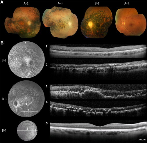
Inherited retinal degenerations, including retinitis pigmentosa (RP) and Leber congenital amaurosis (LCA), comprise a group of disorders showing high genetic and allelic heterogeneity. The determination of a full catalog of genes that can, when mutated, cause human retinal disease is a powerful means to understand the molecular physiology and pathology of the human retina. As more genes are found, remaining ones are likely to be rarer and/or unexpected candidates. Here, we identify a family in which all known RP/LCA-related genes are unlikely to be associated with their disorder. A combination of homozygosity mapping and exome sequencing identifies a homozygous nonsense mutation, c.496C>T (p.Arg166X), in a gene, KCNJ13, encoding a potassium channel subunit Kir7.1. A screen of a further 333 unrelated individuals with recessive retinal degeneration identified an additional proband, homozygous for a missense mutation, c.722T>C (p.Leu241Pro), in the same gene. The three affected members of the two families have been diagnosed with LCA. All have a distinct and unusual retinal appearance and a similar early onset of visual loss, suggesting both impaired retinal development and progressive retinal degeneration, involving both rod and cone pathways. Examination of heterozygotes revealed no ocular disease. This finding implicates Kir7.1 as having an important role in human retinal development and maintenance. This disorder adds to a small diverse group of diseases consequent upon loss or reduced function of inwardly rectifying potassium channels affecting various organs. The distinct retinal phenotype that results from biallelic mutations in KCNJ13 should facilitate the molecular diagnosis in further families.


Figures

Figure 1
Figure 1
Identification of KCNJ13 Mutations in Individuals from Two Families with LCA (A) Pedigree of family A, exome sequencing data from patient A-3, and Sanger sequencing results in affected brother A-2 and unaffected sister A-1. Pedigree reveals a recent common ancestor. Exome sequencing was performed in DNA from patient A-3. An R script assessing and plotting homozygous or heterozygous state of detected SNPs was composed. In chromosome 2, long regions of homozygosity were detected; SNPs within them are represented as red lines. Within the largest region of homozygosity, a nonsense mutation was identified in KCNJ13 (c.496C>T [p.Arg166X]). Gene structure of KCNJ13 is presented (reverse strand), comprising of three exons. Coverage depth distribution of the mapped reads along the three exons is shown (Savant Genome Browser; forward reads are in dark blue, reverse in light blue). Sequencing reads showing the homozygous nonsense mutation are presented (IGV viewer; 80 reads total: 11 forward and 69 reverse, 100% adenine). Electropherograms of DNA sequence surrounding the p.Arg166X mutation are shown for affected brother A-2 and unaffected sister A-1. (B) Pedigree of family B, electropherograms of DNA sequence surrounding the c.722T>C (p.Leu241Pro) mutation in subjects B-3 and B-2, multiple alignment of ten KCNJ13 orthologs around the mutated amino acid, and schematic representation of Kir7.1. The alignment was performed with ClustalW using the following Ensemble transcripts: Homo sapiens ENST00000233826, Pan troglodytes ENSPTRT0000002418, Macaca mulatta ENSMMUT00000030719, Bos taurus ENSBTAT00000007700, Canis familiaris ENSCAFT00000018359, Mus musculus ENSMUST00000113212, Rattus norvegicus ENSRNOT00000021507, Gallus gallus ENSGALT00000002259, Xenopus tropicalis ENSXETT00000030393, and Danio rerio ENSDART00000063777. The schematic representation of Kir7.1 highlights the structural domains, predicted membrane topology, and the location of mutated residue identified.
Figure 2
Figure 2
Retinal Imaging of Patients and Unaffected Family Members Carrying KCNJ13 Mutations (A) Color photographs of right fundi of patient A-2 (aged 34) and patient A-3 (aged 32) in family A as well as left fundus of patient B-3 in family B (aged 33). Mormal left fundus of 39-year old unaffected c.496C>T (p.Arg166X) carrier (A-1, family A) is also shown. (B) Infrared image and linear spectral domain optical coherence tomography scans (SD-OCT; Spectralis HRA+OCT, Heidelberg Engineering, Heidelberg, Germany) of right (1, 2) and left (3, 4) retina of patient B-3 in family B. Right retina of his 64-year-old unaffected mother (B-1) is also shown for comparison (5). On some occasions, image quality was compromised by unstable fixation due to nystagmus or posterior capsule opacification.
Figure 3
Figure 3
Predicted Protein Structure of a Human Kir7.1 Subunit (A) Overlay of wild-type (WT; green) and mutant (yellow) Kir7.1 monomer models. Similar to other Kir family members, the Kir7.1 monomer is comprised of an α-helical membrane domain and an intracellular domain primarily composed of β sheets (asterisk highlights the position of the c.722T>C [p.Leu241Pro]). (B) Detail of the region around the 241 amino acid. Distortion of the protein secondary structure is evident in the mutant when compared to the wild-type monomer. (C and D) The side chains of the Leu241 (wild-type; blue) and Pro241 (mutant; red) residues are highlighted; the cyclic R group structure of proline bends the amino acid chain. (D) presents detail of (C). The structural model was generated by using the SWISS-MODEL protein homology modeling server; the crystal structure of KirBac1.1 (Protein Data Bank code 1q7b) was used as a template. Similar findings were observed when the structure of cKir2.2 (Protein Data Bank code 3jycA) was used as a template. PyMOL (Delano Scientific, Portland, OR) was used to view the three-dimensional molecular structures.

Similar articles

Cited by

References

    1. Wright A.F., Chakarova C.F., Abd El-Aziz M.M., Bhattacharya S.S. Photoreceptor degeneration: genetic and mechanistic dissection of a complex trait. Nat. Rev. Genet. 2010;11:273–284. - PubMed
    1. den Hollander A.I., Roepman R., Koenekoop R.K., Cremers F.P.M. Leber congenital amaurosis: genes, proteins and disease mechanisms. Prog. Retin. Eye Res. 2008;27:391–419. - PubMed
    1. den Hollander A.I., Black A., Bennett J., Cremers F.P. Lighting a candle in the dark: advances in genetics and gene therapy of recessive retinal dystrophies. J. Clin. Invest. 2010;120:3042–3053. - PMC - PubMed
    1. Lander E.S., Botstein D. Homozygosity mapping: a way to map human recessive traits with the DNA of inbred children. Science. 1987;236:1567–1570. - PubMed
    1. Korn J.M., Kuruvilla F.G., McCarroll S.A., Wysoker A., Nemesh J., Cawley S., Hubbell E., Veitch J., Collins P.J., Darvishi K. Integrated genotype calling and association analysis of SNPs, common copy number polymorphisms and rare CNVs. Nat. Genet. 2008;40:1253–1260. - PMC - PubMed

Publication types

MeSH terms