Review
. 2011 Jul 19;21(1):120-33.
doi: 10.1016/j.devcel.2011.06.011.

Planar cell polarity: coordinating morphogenetic cell behaviors with embryonic polarity

Affiliations
Review

Planar cell polarity: coordinating morphogenetic cell behaviors with embryonic polarity

Ryan S Gray et al. Dev Cell. .

Abstract

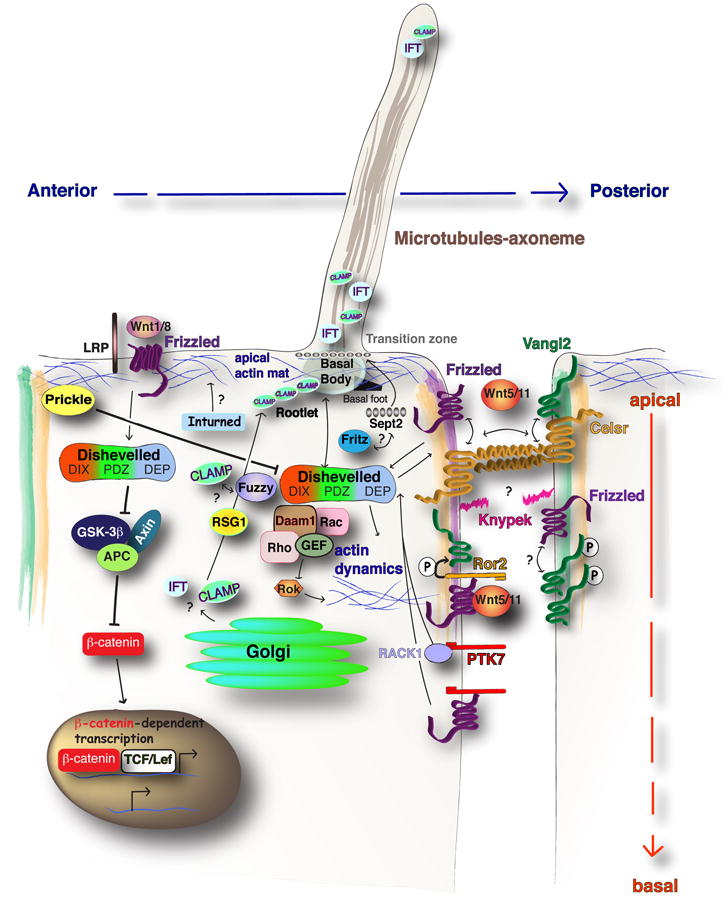
Planar cell polarization entails establishment of cellular asymmetries within the tissue plane. An evolutionarily conserved planar cell polarity (PCP) signaling system employs intra- and intercellular feedback interactions between its core components, including Frizzled, Van Gogh, Flamingo, Prickle, and Dishevelled, to establish their characteristic asymmetric intracellular distributions and coordinate planar polarity of cell populations. By translating global patterning information into asymmetries of cell membranes and intracellular organelles, PCP signaling coordinates morphogenetic behaviors of individual cells and cell populations with the embryonic polarity. In vertebrates, by polarizing cilia in the node/Kupffer's vesicle, PCP signaling links the anteroposterior to left-right embryonic polarity.


Figures

Figure 1
Figure 1. Fz/PCP pathway in epithelial and mesenchymal cell populations
(A) Asymmetric localization of core PCP components in Drosophila epithelial cells. (B) PCP protein localization in polarized and vertebrate mesenchymal cells during C&E cell movements, showing asymmetric localization of Dvl and Pk.
Figure 2
Figure 2. Model for Wnt/β-catenin and Fz/PCP pathway in vertebrates
The divergence of canonical Wnt/β-catenin signaling or non-canonical Fz/PCP signaling relies on Dvl protein. The canonical arm of Dvl activation is downstream of Wnt and may involve Frizzled (Fz) co-receptors such as LRP. This activation serves to inhibit the β-catenin destruction complex of GSK-3β, Axin and APC, leading to β-catenin dependent transcription in the nucleus. Fz/PCP signaling is activated due to the interaction of Wnt/Fz/Dvl complex at the membrane. This can result in activation of effectors of the Actin cytoskeleton. Interactions between apically localized Celsr/Vang and Celsr/Fz heterodimers are proposed to propagate planar polarity between neighboring cells. The PCP co-factors, Glypican4/kynpek, Ptk7 and Ror2 provide additional regulation of PCP signals in vertebrates. A Wnt5a dependent Ror2/Fz interaction promotes phosphorylation of Vang protein, which may promote anterior Vangl accumulation. Parallel mechanisms of Ptk7 can promote membrane recruitment of Dvl through RACK1, or independent of RACK1 via interaction with Fz. Anterior localized Pk inhibits Dvl accumulation at the anterior cell edge. PCP signaling is necessary for posterior localization of the MTOC or basal body structures. The PCP effector genes, Fuz, Intu, and Fritz, support distinct aspects of ciliogenesis.
Figure 3
Figure 3. Coordination of cell behaviors with embryonic polarity during vertebrate development
Representative illustrations of embryos and PCP dependent planar polarized cell behaviors observed during embryonic development. (A) The early segmentation stage zebrafish embryo displays several AP polarized cell behaviors (blue box), including (i) mediolateral intercalation (Jessen et al., 2002), (ii) radial intercalation (Yin et al., 2008), (iii) polarized cell division (Gong et al., 2004), and (iv) anterior directed cell migrations (yellow box) (Yin et al., 2008). (B) The late segmentation (15-somite stage) zebrafish embryo and Kupffer’s vesicle (KV), the LR patterning organ. The basal body (green cylinders) in the KV cells, is positioned near the posterior cell edge (Borovina et al., 2010). (C) The E16.5 mouse embryo displays (i) AP polarity of hair follicles (Guo et al., 2004), (ii) ML polarity of the kinocilium (green circle) and adjacent stereocilia Actin bundles (red circles) (Wang et al., 2006), and (iii) a gradient of Wnt5a orients the proximal-distal (initially oriented along the AP axis) polarity of phosphorylated Vangl2 protein (green shading) and primary cilia/basal body (red circle) in chondrocytes of the mouse limb (Gao et al., 2011).

References

    1. Adler PN. Planar signaling and morphogenesis in Drosophila. Dev Cell. 2002;2:525–535. - PubMed
    1. Adler PN, Charlton J, Liu J. Mutations in the cadherin superfamily member gene dachsous cause a tissue polarity phenotype by altering frizzled signaling. Development. 1998;125:959–968. - PubMed
    1. Adler PN, Krasnow RE, Liu J. Tissue polarity points from cells that have higher Frizzled levels towards cells that have lower Frizzled levels. Curr Biol. 1997;7:940–949. - PubMed
    1. Aigouy B, Farhadifar R, Staple DB, Sagner A, Roper JC, Julicher F, Eaton S. Cell flow reorients the axis of planar polarity in the wing epithelium of Drosophila. Cell. 2010;142:773–786. - PubMed
    1. Almuedo-Castillo M, Salo E, Adell T. Dishevelled is essential for neural connectivity and planar cell polarity in planarians. Proc Natl Acad Sci U S A. 2011;108:2813–2818. - PMC - PubMed

Publication types

Substances

LinkOut - more resources