Review
. 2011 Jul 19;21(1):172-85.
doi: 10.1016/j.devcel.2011.06.016.

The cellular basis for animal regeneration

Affiliations
Review

The cellular basis for animal regeneration

Elly M Tanaka et al. Dev Cell. .

Abstract

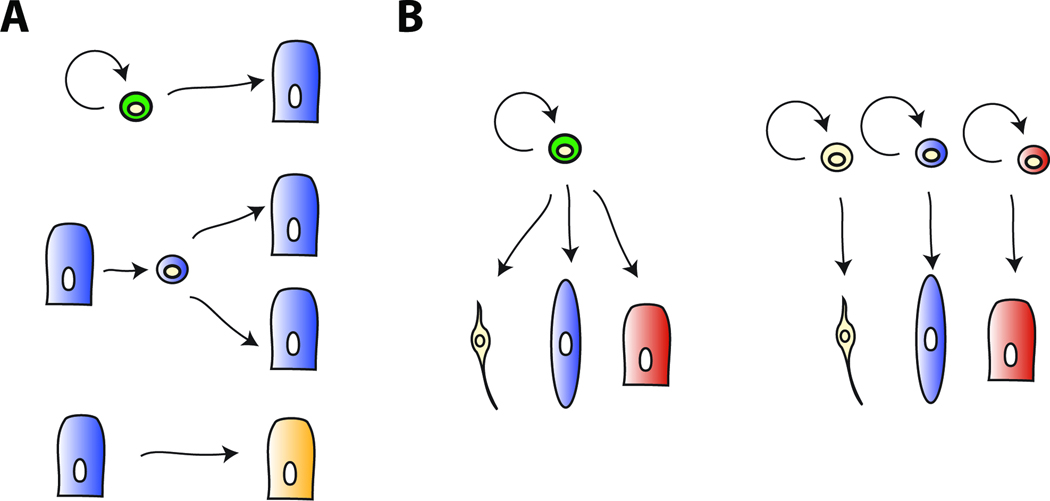
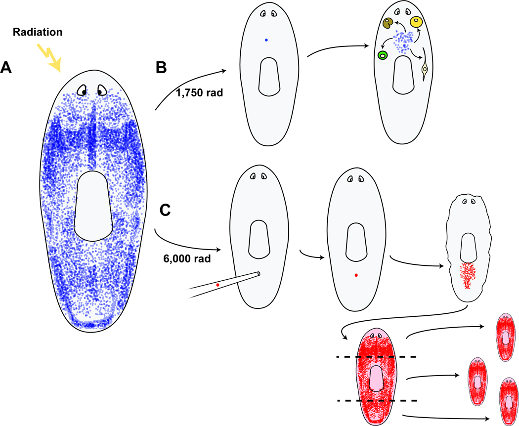
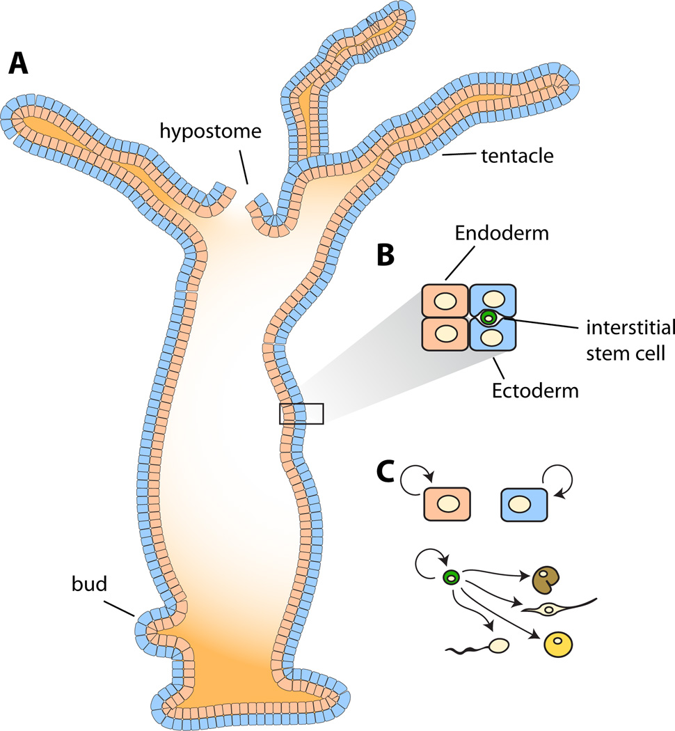
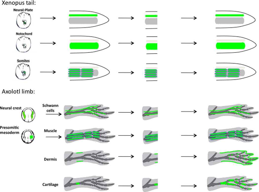
The ability of animals to regenerate missing parts is a dramatic and poorly understood aspect of biology. The sources of new cells for these regenerative phenomena have been sought for decades. Recent advances involving cell fate tracking in complex tissues have shed new light on the cellular underpinnings of regeneration in Hydra, planarians, zebrafish, Xenopus, and Axolotl. Planarians accomplish regeneration with use of adult pluripotent stem cells, whereas several vertebrates utilize a collection of lineage-restricted progenitors from different tissues. Together, an array of cellular strategies-from pluripotent stem cells to tissue-specific stem cells and dedifferentiation-are utilized for regeneration.


Figures

Figure 1
Figure 1. Sources for new cells in regeneration
A. Top, stem cells self-renew and produce one or more differentiated cells. Middle, dedifferentiation is the process by which a cell loses differentiated character to produce a progenitor cell that can divide to produce more differentiated cells. Bottom, transdifferentiation involves the change of one cell type into others. This could occur without division, or following de-differentiation of one cell type into a progenitor for additional cell types. B. Distinct ways for accomplishing tissue-level pluripotency. Left, a pluripotent progenitor cell (a stem cell is depicted) produces differentiated progeny cells spanning multiple germ layers. There could exist multiple, and/or self-renewing intermediates along different lineage paths. Right, different lineage-restricted progenitor cells (stem cell types are depicted) each produce different differentiated cells. Each different tissue separately generates or harbors a restricted stem cell. These stem cells together can reconstitute the three different tissues, while any individual on its own is not sufficient.
Figure 2
Figure 2. Planarian regeneration is accomplished with pluripotent stem cells called cNeoblasts
A. Neoblasts (blue), are the somatic dividing cells of planarians and are depicted in blue. Neoblasts are scattered throughout the body, but restricted to behind the eyes and absent from the pharynx (centrally located). B. Irradiation with 1,750 rad can result in animals with a single surviving neoblast. This single cell can divide and produce a colony of neoblasts, ultimately producing differentiated cells spanning germ layers (Wagner et al., 2011). For example, individual neoblasts can generate both neurons and intestine cells, as well as defined neoblast progeny populations. C. Irradiation with 6,000 rad eliminates all neoblasts. Transplant of a single cNeoblast from a donor strain (red) results in clonogenic growth and, ultimately, the restored capacity for regeneration.
Figure 3
Figure 3. Hydra regeneration is accomplished with three different stem cell populations
A. Hydra are cnidarians with a primary body axis containing a hypostome (or head) at one end and a foot at the other. Cell proliferation in the body column continually pushes cells to the poles of the body. Asexual reproduction is accomplished by budding. B. The body wall contains two epithelial cell layers, ectodermal and endodermal epithelial cells. Interstitial stem cells exist within the ectodermal epithelial cell layer. C. The ectodermal and endodermal epithelial cells proliferate continuously to maintain these tissue layers, producing differentiated epithelial cells, and are therefore considered to be distinct stem cells. A third stem cell type, the multipotent, interstitial stem cell can self-renew and produce neurons, nematocytes, secretory cells, and gametes.
Figure 4
Figure 4. Cell tracking of GFP-labeled cells in amphibians shows that vertebrate appendage regeneration occurs by producing lineage-restricted progenitors in the Xenopus tail and Axolotl limb blastema
Cell labeling was primarily achieved via grafting of embryonic tissues during the neurula stage from GFP-expressing donors to normal hosts. (Top) Xenopus: posterior neural plate, presomitic mesoderm, and notochord were transplanted to label tail spinal cord, muscle, and notochord, respectively. After tail amputation, the labeled tissues regenerated the same tissue type as prior to amputation (Gargioli and Slack, 2004). (Bottom) Axolotl limb Schwann cells and muscle were labeled by embryonic presomitic mesoderm and neural crest transplantation (Kragl et al., 2009). Dermis and cartilage were labeled by direct tissue transplantation in the limb, as well as embryonic tissue grafts (Kragl et al., 2009). After limb amputation, labeled Schwann cells regenerated Schwann cells only. Muscle regenerated muscle and no cartilage. Dermis regenerated dermis, cartilage and connective tissues (also described by Dunis and Namenwirth, 1977), while cartilage regenerated cartilage (also described by Steen, 1968).
Figure 5
Figure 5. Cre/loxP-based cell fate mapping establishes that dedifferentiation occurs during zebrafish heart and fin regeneration
A. Prior to heart resection, cardiomyocytes were labeled via a cardiomyocyte-specific promoter driving the CreER sequence. CreER acted on a cardiomyocyte-specific loxP reporter where a floxed STOP cassette was excised by CreER, which is active in the presence of 4-HT (4-hydroxytamoxifen), to induce GFP expression. Newly regenerated cardiomyocytes (right, below dotted line) express GFP, indicating that they derived from cardiomyocytes in the injured heart tissue (Kikuchi et al, 2010; Jopling et al, 2010). B. Tracking of osteoblasts during caudal fin regeneration demonstrates that they contribute to the regenerated fin and remain restricted to an osteoblast identity. osterix:Cre-ERT2 acting on the loxP reporter; upon Cre-mediated excision of a STOP cassette, the hsp70 promoter drives expression of GFP. GFP expression was induced prior to fin amputation leading to sporadic cell labeling. GFP-expressing cells generate osteoblasts in the regenerated fin, indicating that osteoblasts de-differentiated and divided to produce more osteoblasts, remaining restricted to the osteoblast fate during regeneration (Knopf et al, 2011).

Similar articles

Cited by

References

    1. Aboobaker AA. Planarian stem cells: a simple paradigm for regeneration. Trends Cell Biol. 2011 - PubMed
    1. Adell T, Cebria F, Saló E. Gradients in planarian regeneration and homeostasis. Cold Spring Harb Perspect Biol. 2010;2 a000505. - PMC - PubMed
    1. Adell T, Saló E, Boutros M, Bartscherer K. Smed-Evi/Wntless is required for beta-catenin-dependent and -independent processes during planarian regeneration. Development. 2009;136:905–910. - PubMed
    1. Adoutte A, Balavoine G, Lartillot N, Lespinet O, Prud'homme B, de Rosa R. The new animal phylogeny: reliability and implications. Proc Natl Acad Sci USA. 2000;97:4453–4456. - PMC - PubMed
    1. Akimenko MA, Mari-Beffa M, Becerra J, Geraudie J. Old questions, new tools, and some answers to the mystery of fin regeneration. Dev Dyn. 2003;226:190–201. - PubMed

MeSH terms

LinkOut - more resources