. 2011 Sep;179(3):1542-8.
doi: 10.1016/j.ajpath.2011.05.049. Epub 2011 Jul 19.

Prolonged infusion of angiotensin II in apoE(-/-) mice promotes macrophage recruitment with continued expansion of abdominal aortic aneurysm

Affiliations

Prolonged infusion of angiotensin II in apoE(-/-) mice promotes macrophage recruitment with continued expansion of abdominal aortic aneurysm

Debra L Rateri et al. Am J Pathol. 2011 Sep.

Abstract

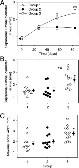
Angiotensin II (AngII) infusion initiates abdominal aortic aneurysm (AAA) development due to medial disruption and results in luminal dilation and thrombus formation. The objective of this study was to determine whether AAA progressed during protracted AngII infusion. Male apoE(-/-) mice were infused with AngII using miniosmotic pumps. On day 27, suprarenal aortic luminal diameters were ultrasonically measured to identify mice exhibiting AAAs. Mice were designated to three groups with similar mean luminal dilation. Group 1 mice were sacrificed on day 28. Group 2 and 3 mice were subsequently infused with saline or AngII, respectively, for an additional 56 days. In Group 2, saline infusion-after the initial 28 days of AngII infusion-led to an immediate decrease in systolic blood pressure. Over the subsequent 56 days of saline infusion, there were no aneurysm-related deaths or significant changes in luminal diameter. In contrast, continuous AngII infusion in Group 3 maintained persistently increased systolic blood pressure, with aneurysmal rupture-associated deaths, increased luminal diameters, and tissue remodeling. Aortic aneurysmal segments that expanded during continuous AngII infusion exhibited macrophage accumulation in regions of medial disruption, predominantly on the adventitial aspect. Macrophages immunostained for CD206 more than for iNOS, consistent with an M2 phenotype. In conclusion, prolonged AngII infusion promotes AAA expansion, and is associated with enhanced rupture rates and increased macrophage infiltration.


Figures

Figure 1
Figure 1
Systolic blood pressure and mortality during prolonged infusion of either AngII or saline. A: Systolic blood pressure was measured continuously for 5 days per week at the indicated intervals. Means of groups are represented by symbols and SEM by bars. *P < 0.0001 for comparison of Groups 2 and 3 during the 28- to 84-day interval using a linear mixed model. B: Symbols represent the number of surviving mice during the 56-day infusion of either AngII or saline.
Figure 2
Figure 2
In vivo and ex vivo dimensions of suprarenal aortas measured in mice infused with either AngII or saline. A: Luminal diameters of the suprarenal aortas were measured in vivo ultrasonically before AngII infusion and at 28-day intervals. Symbols represent means, and bars are SEM *P = 0.0014, **P < 0.0001 for comparisons of Groups 2 and 3 using a linear mixed model. B: Individual suprarenal aortic diameters, measured in vivo ultrasonically, are represented by triangles and circles, mean data are represented by diamonds, and SEM are represented by bars. *P < 0.001 for comparisons of Group 3 to Groups 1 and 2 by analysis of variance followed by post hoc pairwise multiple comparison procedures using the Holm-Sidak method. C: Individual suprarenal aortic widths, measured ex vivo, are represented by triangles and circles, mean data are represented by diamonds, and SEMs are represented by bars. *P = 0.004 for comparisons of Group 3 to Groups 1 and 2 using Kruskal-Wallis analysis of variance on ranks followed by post hoc multiple comparison procedures using the nonparametric Dunn's method.
Figure 3
Figure 3
Examples of suprarenal aortas of apoE−/− mice infused with different intervals of AngII (1000 ng/kg/min). After 28 days of AngII infusion, the suprarenal aorta is dilated and frequently contains overt thrombotic material. After 84 days of AngII infusion, the suprarenal aorta can be greatly dilated (over 4 mm from a baseline of 0.8 mm) with thinned wall and resolution of thrombotic material. To facilitate cryosectioning of these thinned and dilated tissues, they are routinely perfusion fixed and filled with low melting point agarose containing green dye to assist visualization of the tissue in the optimal cutting temperature compound.
Figure 4
Figure 4
Movat's pentachrome staining of an entire abdominal aortic aneurysm. The full length of an AAA was serial sectioned, and every tenth slide was stained with Movat's pentachrome. Section (A, B, and C) starts proximally to the AAA and sections continue through it (D and E), finishing distally to the dilation (F). Arrows point to elastin breaks (D and E). T, thrombus. Original magnification, 400×.
Figure 5
Figure 5
Macrophage counting throughout the suprarenal aneurysmal aorta. A: The mean number of macrophages per section of all samples in each group. Histobars represent means, and bars represent SEM. *P = 0.0005 for comparison of Group 3 to Group 1; P = 0.0002 for comparison of Group 3 to 2 using a linear mixed model. B: The mean number of macrophages located in the adventitia of all samples in each group. Histobars represent means, and bars represent SEM, *P = 0.02, P = 0.002 for comparisons of Group 3 to Groups 1 and 2, respectively, using a linear mixed model.
Figure 6
Figure 6
Macrophage counting and medial breakage throughout the suprarenal aneurysmal aorta. Examples (three per group, A, B, and C) of macrophage numbers per section throughout the aneurysmal suprarenal aorta of all of the three groups described in this study. Symbols represent macrophage counts per section. Shaded areas represent regions of elastin breaks associated with luminal dilation.

Similar articles

Cited by

References

    1. Daugherty A., Cassis L. Chronic angiotensin II infusion promotes atherogenesis in low density lipoprotein receptor −/− mice. Ann N Y Acad Sci. 1999;892:108–118. - PubMed
    1. Daugherty A., Manning M.W., Cassis L.A. Angiotensin II promotes atherosclerotic lesions and aneurysms in apolipoprotein E-deficient mice. J Clin Invest. 2000;105:1605–1612. - PMC - PubMed
    1. Wang Y.X., Martin-McNulty B., Freay A.D., Sukovich D.A., Halks-Miller M., Li W.W., Vergona R., Sullivan M.E., Morser J., Dole W.P., Deng G.G. Angiotensin II increases urokinase-type plasminogen activator expression and induces aneurysm in the abdominal aorta of apolipoprotein E-deficient mice. Am J Pathol. 2001;159:1455–1464. - PMC - PubMed
    1. Bruemmer D., Collins A.R., Noh G., Wang W., Territo M., Arias-Magallona S., Fishbein M.C., Blaschke F., Kintscher U., Graf K., Law R.E., Hsueh W.A. Angiotensin II-accelerated atherosclerosis and aneurysm formation is attenuated in osteopontin-deficient mice. J Clin Invest. 2003;112:1318–1331. - PMC - PubMed
    1. Ishibashi M., Egashira K., Zhao Q., Hiasa K., Ohtani K., Ihara Y., Charo I.F., Kura S., Tsuzuki T., Takeshita A., Sunagawa K. Bone marrow-derived monocyte chemoattractant protein-1 receptor CCR2 is critical in angiotensin II-induced acceleration of atherosclerosis and aneurysm formation in hypercholesterolemic mice. Arterioscler Thromb Vasc Biol. 2004;24:174–178. - PubMed

Publication types

MeSH terms