. 2011 Sep;179(3):1265-77.
doi: 10.1016/j.ajpath.2011.05.042. Epub 2011 Jul 19.

Minocycline attenuates photoreceptor degeneration in a mouse model of subretinal hemorrhage microglial: inhibition as a potential therapeutic strategy

Affiliations

Minocycline attenuates photoreceptor degeneration in a mouse model of subretinal hemorrhage microglial: inhibition as a potential therapeutic strategy

Lian Zhao et al. Am J Pathol. 2011 Sep.

Abstract

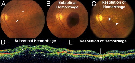
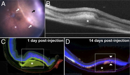
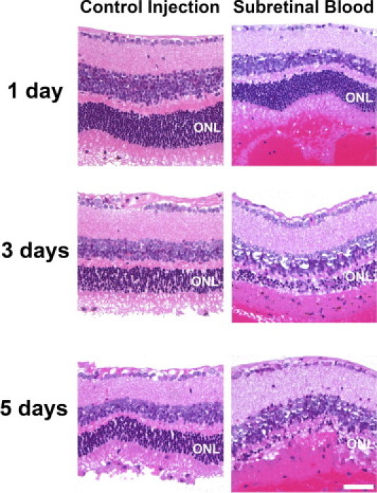
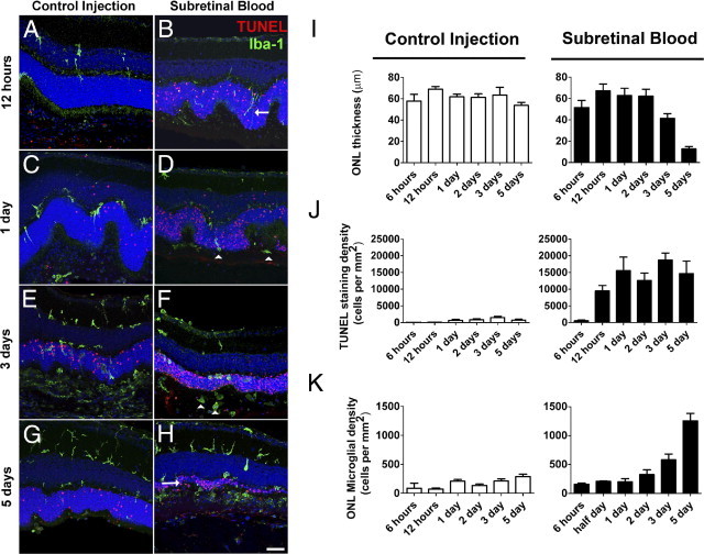
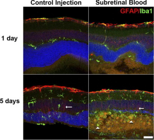
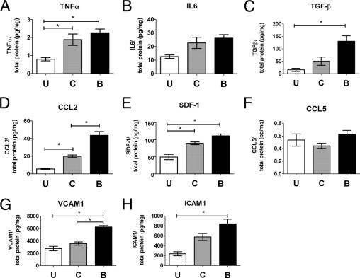
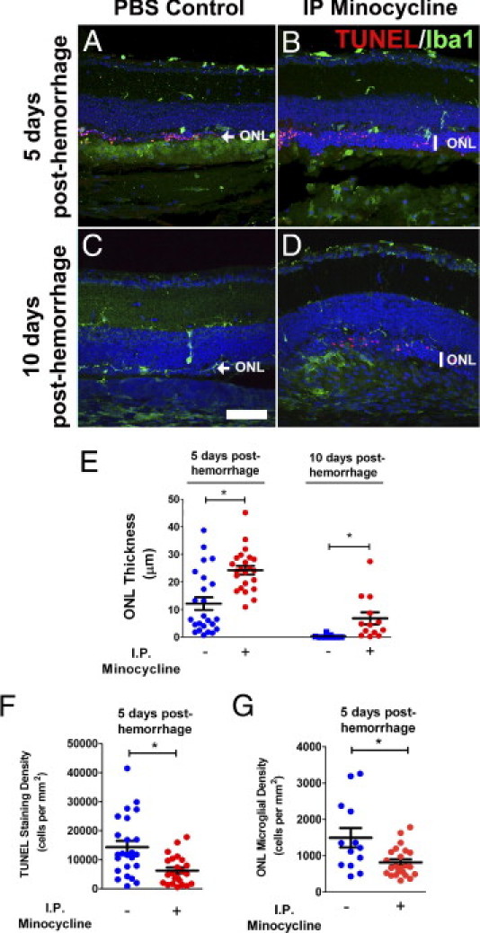
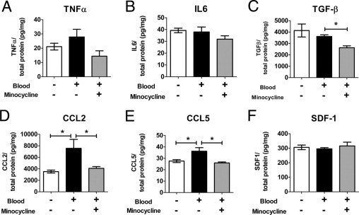
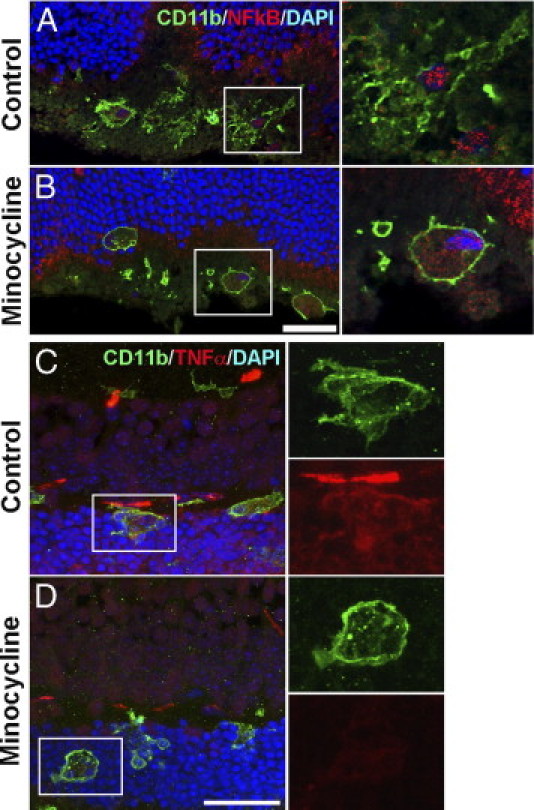
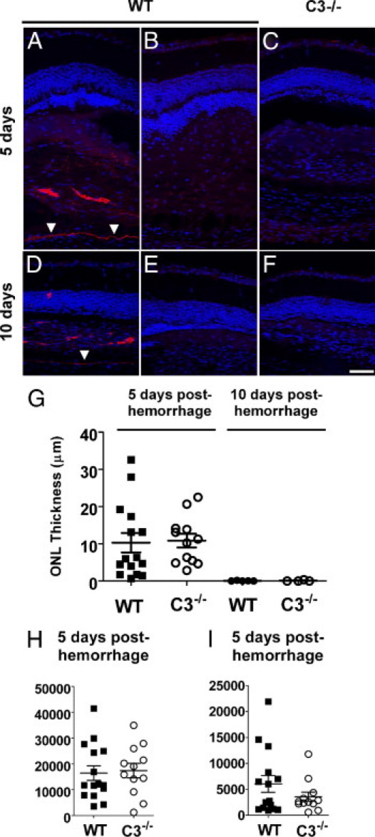
Hemorrhage under the neural retina (subretinal hemorrhage) can occur in the context of age-related macular degeneration and induce subsequent photoreceptor cell death and permanent vision loss. Current treatments with the objective of removing or displacing the hemorrhage are invasive and of mixed efficacy. We created a mouse model of subretinal hemorrhage to characterize the inflammatory responses and photoreceptor degeneration that occur in the acute aftermath of hemorrhage. It was observed that microglial infiltration into the outer retina commences as early as 6 hours after hemorrhage. Inflammatory cells progressively accumulate in the outer nuclear layer concurrently with photoreceptor degeneration and apoptosis. Administration of minocycline, an inhibitor of microglial activation, decreased microglial expression of chemotactic cytokines in vitro and reduced microglial infiltration and photoreceptor cell loss after subretinal hemorrhage in vivo. Inflammatory responses and photoreceptor atrophy occurred after subretinal hemorrhage, however, the degree of response and atrophy were similar between C3-deficient and C3-sufficient mice, indicating a limited role for complement-mediated processes. Our data indicate a role for inflammatory responses in driving photoreceptor cell loss in subretinal hemorrhage, and it is proposed that microglial inhibition may be beneficial in the treatment of subretinal hemorrhage.


Figures

Figure 1
Figure 1
Clinical findings in subretinal hemorrhage in the context of neovascular (wet) AMD. Fundus photographs demonstrate the typical clinical setting in which subretinal hemorrhage develops in the context of AMD. A: Subretinal hemorrhage can occur acutely in an eye at risk such as one containing signs of intermediate AMD in the form of drusen and pigmentary changes (arrows). B: Appearance of an acute macular subretinal hemorrhage as a result of choroidal neovascularization formation. C: Subretinal blood gradually resolves over a period of weeks, with the affected area developing extensive retinal atrophy and loss of central vision (arrowheads). D: Corresponding images obtained using OCT in the same eye demonstrate in vivo cross-sectional views of the subretinal hemorrhage that detaches the retina at the macula (arrowheads) in a dome-shaped configuration. E: With resolution of the hemorrhage, concurrent retinal atrophy and loss of the outer retinal lamina in the affected area is typically observed (arrows).
Figure 2
Figure 2
Anatomical and pathologic findings in a murine model of subretinal hemorrhage. A model for subretinal hemorrhage was created in the adult mouse by injecting a small volume (1.6 μL) of autologous blood into the subretinal space via a transscleral route using a small-gauge (33G) needle. A:In vivo photograph of the mouse fundus of the treated eye at 5 hours after subretinal blood injection reveals an area of dome-shaped retinal detachment (white arrows) containing a central blood clot (black arrow). B:In vivo cross-sectional view of the subretinal hemorrhage (asterisk) at 3 days after treatment as visualized using OCT. C: Histologic section of the retina in the area of hemorrhage reveals an area of retinal detachment (box) overlying subretinal hemorrhage (asterisk). Photoreceptor apoptosis in the area overlying the hemorrhage is labeled using TUNEL (red). Nuclear staining was performed using DAPI (blue), and the green channel was used to demonstrate the autofluorescent nature of the subretinal hemorrhage. D: Histologic section of the retina at 2 weeks after subretinal blood injection demonstrates resolution of the hemorrhage (asterisk) and development of outer retinal atrophy over the area of hemorrhage (arrowheads).
Figure 3
Figure 3
Outer retinal atrophy after subretinal hemorrhage in a mouse model of subretinal hemorrhage. Experimental mice received a subretinal injection of autologous blood (1.6 μL) in the treated eye, and in the contralateral control eye, a subretinal injection of an equal volume of an inert viscoelastic substance (0.5% sodium hyaluronate) to simulate an equivalent retinal detachment without the presence of subretinal blood. Representative photomicrographs of thin plastic retinal sections stained using H&E are shown at 1, 3, and 5 days after injection. Outer nuclear layer thickness (ONL) was reduced in eyes receiving subretinal blood injections (right panels) to a significantly greater extent than in eyes receiving control injections (left panels). Scale bar = 50 μm.
Figure 4
Figure 4
Photoreceptor apoptosis and microglial activation after subretinal hemorrhage. Experimental animals received subretinal injections of either blood or viscoelastic substance (0.5% sodium hyaluronate; control). Eyes were harvested for histopathologic analysis at various times after injection, and were analyzed using TUNEL staining (red) or immunohistochemistry for Iba-1 (to label microglia, green). A and B: At 12 hours after injection, retinal microglia, which are normally confined to the inner retina, could be seen extending vertically oriented processes and migrating into the outer nuclear layer in eyes injected with subretinal blood (B, arrow) but to a smaller extent in control eyes (A). TUNEL positivity is observed in the outer nuclear layer in eye injected with subretinal blood but was largely absent in control eyes. C and D: At 1 day after injection, the number of TUNEL-positive cells increased in both control eyes (C) and eyes injected with subretinal blood eyes (D) but remained significantly higher in eyes injected with subretinal blood. Microglia were also observed to migrate into the outer retina, translocating their somata into the outer nuclear layer and subretinal space (arrowheads). E and F: At 3 days, the thickness of the outer nuclear layer was considerably decreased, to a greater extent in eyes injected with subretinal blood (F) than in control eyes (E). F: Microglia in the outer nuclear layer and subretinal space also acquired a rounded amoeboid architecture in eyes injected with subretinal blood (arrowheads). G and H: At 5 days, the outer nuclear layer in eyes injected with subretinal blood was reduced to only one or two cells thick (H, arrow) but remained substantial in control eyes (G). I: Quantification of outer retinal layer thickness, photoreceptor apoptosis, and outer retinal microglial infiltration after control and subretinal blood injections demonstrated that outer nuclear layer thickness was largely maintained over 5 days after injection in control eyes (left), whereas after subretinal blood injection, it decreased significantly at 3 and 5 days after injection (right). J: Density of TUNEL-positive cells in the outer nuclear layer remained relatively low in control eyes (left), whereas those in eyes injected with subretinal blood increased markedly at 12 hours (right). K: Density of microglial infiltration into the outer nuclear layer increased over time, and was relatively higher at 3 and 5 days after subretinal blood injection compared with control injection. Scale bar = 50 μm.
Figure 5
Figure 5
Microglia and Müller cell gliosis in mouse model of subretinal hemorrhage. Microglia gliosis and Müller cell gliosis were followed up at 1 and 5 days after injection with immunohistochemical staining for Iba-1 (green) to label microglia and GFAP (red), astrocytes, and activated Müller cells. At 1 day after injection, weak GFAP immunopositivity in Müller cell processes was observed in both control eyes and eyes injected with subretinal blood. At 5 days after injection, Müller cell processes (arrows) were clearly labeled with GFAP in both control eyes and those that received subretinal injections, indicating an increase in Müller cell gliosis. Large numbers of microglia with prominent, rounded, amoeboid architecture were apparent in the outer nuclear layer and subretinal space (arrowheads), indicating a more extensive degree of microgliosis in eyes injected with subretinal blood compared with control eyes injected with viscoelastic material. Scale bar = 50 μm.
Figure 6
Figure 6
Inflammatory cytokines, chemotactic cytokines, and adhesion molecules are up-regulated after subretinal hemorrhage. Retinal levels of cytokines and adhesion molecules in the retina were assessed using ELISA in uninjected eyes (U) and in eyes injected subretinally 3 days before with either control (C) or autologous blood (B). Inflammatory cytokines TNF-α (A), IL-6 (B), and transforming growth factor-β (C) were generally elevated in injected eyes compared with uninjected eyes, with the highest levels in eyes injected with autologous blood. Similar trends were observed for chemotactic cytokines CCL2 (D) and SDF-1 (E) and adhesion molecules VCAM-1 (G) and ICAM-1 (H). Levels of chemokine CCL5 (F) were low and relatively unchanged. *P < 0.05).
Figure 7
Figure 7
Minocycline treatment reduces outer nuclear layer apoptosis and atrophy after subretinal injection. Experimental animals were treated with minocycline (i.p. injection, 50 mg/kg b.i.d.) beginning immediately after injection of subretinal blood. As control, experimental animals were injected with PBS at the same schedule after subretinal blood injection. Representative retinal sections comparing atrophy in the outer nuclear layer (ONL) in PBS-treated control animals and minocycline-treated animals at 5 days (A and B) and 10 days (C and D) after injection of subretinal blood (TUNEL staining in red, and Iba-1 staining in green). At 5 days after injection, outer nuclear layer thickness was typically greater in minocycline-treated animals than in control-treated animals. Density of TUNEL-positive cells and microglia in the outer retina were correspondingly lower in minocycline-treated animals. At 10 days after injection, outer nuclear layer thickness in control retinas was either completely attenuated or reduced to a layer one cell thick (arrow). Outer nuclear layer thickness was significantly greater in minocycline-treated animals (vertical bar). E: Quantitation of outer nuclear layer thickness in eyes from control (−) and minocycline-treated (+) animals at 5 and 10 days after hemorrhage. F: Density of TUNEL-positive cells in the outer nuclear layer in control (−) and minocycline-treated (+) animals at 5 days after subretinal injection. G: Density of microglial cells in the outer nuclear layer in control (−) and minocycline-treated (+) animals at 5 days after subretinal injection. Data for postinjection day 5: n = 24 eyes from 12 control animals and 23 eyes from 12 treated animals. Data for postinjection day 10: n = 9 eyes from 6 control animals and 13 eyes from 7 treated animals. *P < 0.05). Scale bar = 50 μm.
Figure 8
Figure 8
Effect of minocycline on expression of cytokines by retinal microglia after exposure to red blood cells. Levels of cytokines in cultured retinal microglia were assessed using ELISA under baseline conditions (white bars), after exposure to lysed red blood cells (black bars), and after exposure to lysed red blood cells and minocycline together (gray bars). A: TNF-α. B: IL-6. C: Transforming growth factor-β. D: CCL2. E: CCL5. F: SDF-1. *P < 0.05).
Figure 9
Figure 9
Effect of minocycline on microglial activation in the retina after subretinal hemorrhage. Subretinal hemorrhage was induced in experimental animals, and treated daily with i.p. injection of minocycline or PBS. Retinal tissue was isolated after 3 days of treatment. A and B: Microglia in the outer retina were labeled with CD11b (green), and their nuclei were marked using DAPI staining. Cellular localization of NF-κB (subunit p65) was followed by immunohistochemical staining (red). A: Microglia in PBS-treated control retina demonstrated a pattern of NF-κB staining that was primarily confined to the nucleus, suggestive of NF-κB activation. B: Microglia in minocycline-treated retina demonstrated a more diffuse cytoplasmic pattern of NF-κB staining, indicative of a less-activated state. C and D: CD11b-positive microglia (green) in the outer retina were co-labeled with an antibody to TNF-α (red). Microglia in PBS-treated control retinas (C) demonstrated a higher level of TNF-α immunopositivity than did those in minocycline-treated retinas (D), indicating a suppressive effect on microglial TNF- α expression by minocycline. Representative images shown were captured using confocal microscopy under similar settings to provide a relative comparison of expression levels. Scale bar = 30 μm.
Figure 10
Figure 10
Role of complement in outer nuclear layer atrophy in a mouse model of subretinal blood. Immunohistochemical staining for C3 after subretinal injection demonstrated C3 deposition in the subretinal space and along Bruch's membrane. Representative retinal sections demonstrated C3 deposition in a wild-type C57BL/6 mouse at 5 days (A) and 10 days (D) after injection of subretinal blood. Absence of C3 staining was observed in control experiments in which the primary antibody to C3 was omitted (B and E) and when similar sections from a C3−/− mouse on a C57Bl6 background were tested (C and F). Arrowheads indicate the position of Bruch's membrane. G: Quantitation of outer nuclear layer thickness in eyes from wild-type and C3−/− animals at 5 and 10 days after injection of subretinal blood. H: Density of TUNEL-positive cells in the outer nuclear layer in wild-type and C3−/− animals at 5 days after subretinal injection. I: Density of microglial cells in wild-type and C3−/− animals at 5 days after subretinal injection. No significant differences were detected (P > 0.05) for all comparisons between wild-type and C3−/− animals. Data for postinjection day 5: n = 15 eyes from 8 wild-type animals and 12 eyes from 6 C3−/− animals. Data for postinjection day 10: n = 5 eyes from 3 wild-type animals and 4 eyes from 3 C3−/− animals. Scale bar = 50 μm.

References

    1. Congdon N., O'Colmain B., Klaver C.C., Klein R., Munoz B., Friedman D.S., Kempen J., Taylor H.R., Mitchell P. Causes and prevalence of visual impairment among adults in the United States. Arch Ophthalmol. 2004;122:477–485. - PubMed
    1. Rein D.B., Wittenborn J.S., Zhang X., Honeycutt A.A., Lesesne S.B., Saaddine J. Forecasting age-related macular degeneration through the year 2050: the potential impact of new treatments. Arch Ophthalmol. 2009;127:533–540. - PubMed
    1. Kawasaki R., Yasuda M., Song S.J., Chen S.J., Jonas J.B., Wang J.J., Mitchell P., Wong T.Y. The prevalence of age-related macular degeneration in Asians: a systematic review and meta-analysis. Ophthalmology. 2010;117:921–927. - PubMed
    1. Mousa S.A., Mousa S.S. Current status of vascular endothelial growth factor inhibition in age-related macular degeneration. BioDrugs. 2010;24:183–194. - PubMed
    1. Bressler N.M. Antiangiogenic approaches to age-related macular degeneration today. Ophthalmology. 2009;116:S15–S23. - PubMed

Publication types

MeSH terms