

A role for vascular deficiency in retinal pathology in a mouse model of ataxia-telangiectasia
- PMID: 21763675
- PMCID: PMC3157193
- DOI: 10.1016/j.ajpath.2011.05.026
A role for vascular deficiency in retinal pathology in a mouse model of ataxia-telangiectasia
Abstract
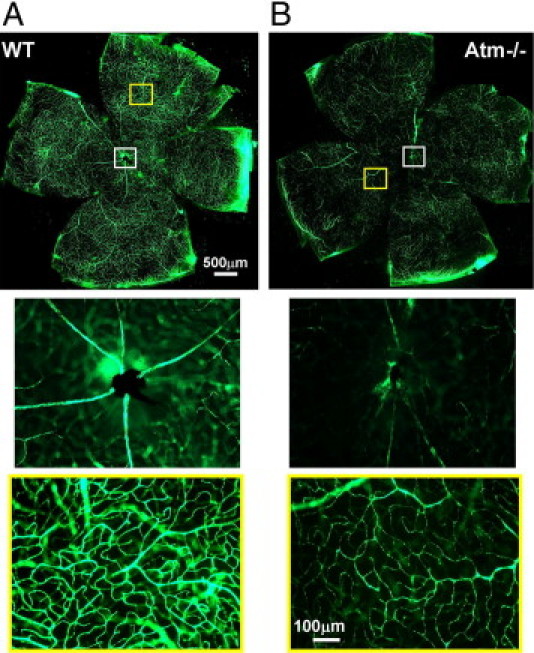
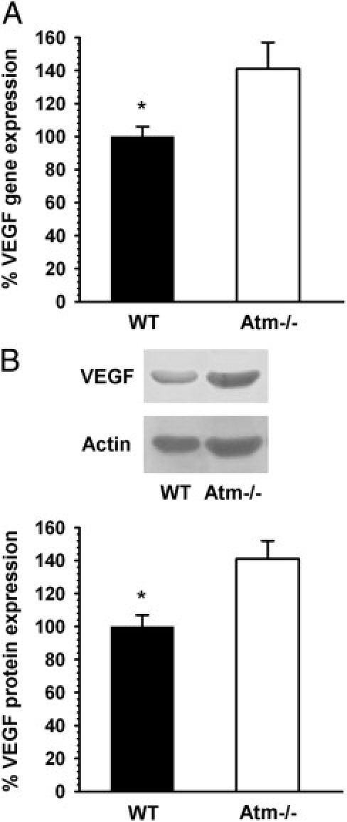
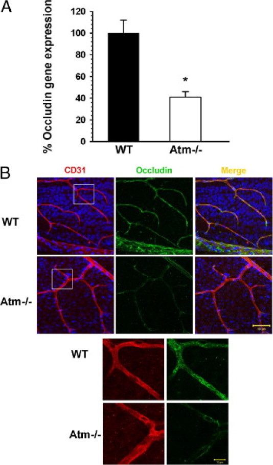
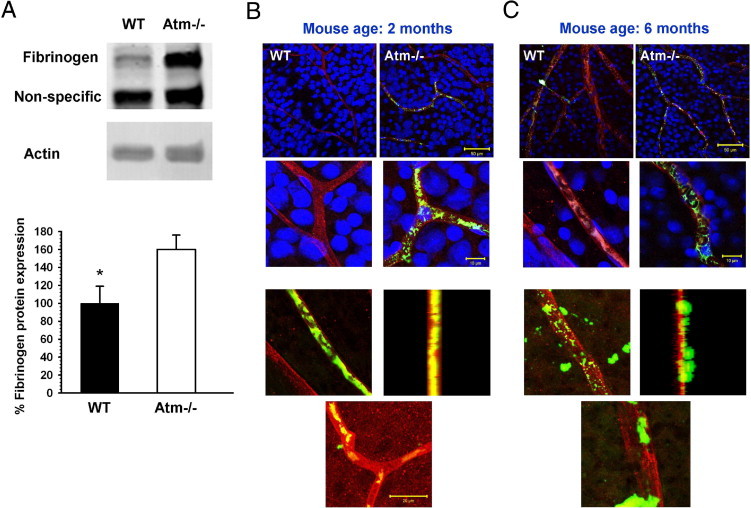
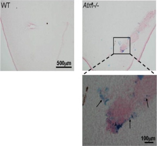
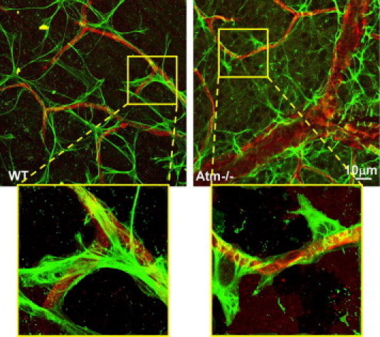
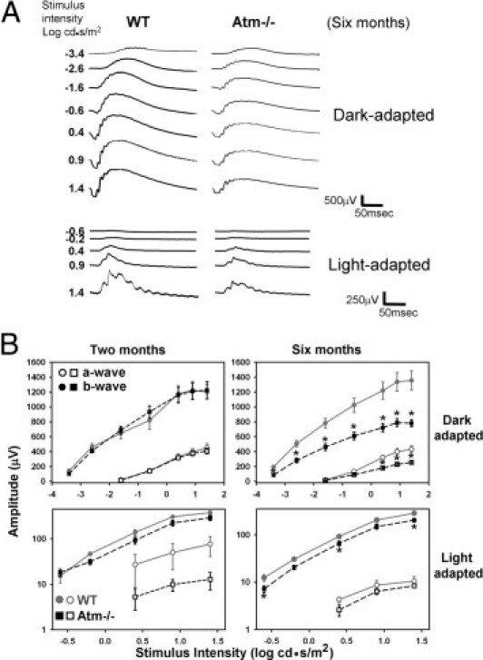
Ataxia-telangiectasia is a multifaceted syndrome caused by null mutations in the ATM gene, which encodes the protein kinase ATM, a key participant in the DNA damage response. Retinal neurons are highly susceptible to DNA damage because they are terminally differentiated and have the highest metabolic activity in the central nervous system. In this study, we characterized the retina in young and aged Atm-deficient mice (Atm(-/-)). At 2 months of age, angiography revealed faint retinal vasculature in Atm(-/-) animals relative to wild-type controls. This finding was accompanied by increased expression of vascular endothelial growth factor protein and mRNA. Fibrinogen, generally absent from wild-type retinal tissue, was evident in Atm(-/-) retinas, whereas mRNA of the tight junction protein occludin was significantly decreased. Immunohistochemistry labeling for occludin in 6-month-old mice showed that this decrease persists in advanced stages of the disease. Concurrently, we noticed vascular leakage in Atm(-/-) retinas. Labeling for glial fibrillary acidic protein demonstrated morphological alterations in glial cells in Atm(-/-) retinas. Electroretinographic examination revealed amplitude aberrations in 2-month-old Atm(-/-) mice, which progressed to significant functional deficits in the older mice. These results suggest that impaired vascularization and astrocyte-endothelial cell interactions in the central nervous system play an important role in the etiology of ataxia-telangiectasia and that vascular abnormalities may underlie or aggravate neurodegeneration.
Copyright © 2011 American Society for Investigative Pathology. Published by Elsevier Inc. All rights reserved.
Figures
Similar articles
-
Alterations of retinal vasculature in cystathionine-Beta-synthase mutant mice, a model of hyperhomocysteinemia.Invest Ophthalmol Vis Sci. 2013 Feb 1;54(2):939-49. doi: 10.1167/iovs.12-10536. Invest Ophthalmol Vis Sci. 2013. PMID: 23307965 Free PMC article.
-
Altered expression of retinal occludin and glial fibrillary acidic protein in experimental diabetes. The Penn State Retina Research Group.Invest Ophthalmol Vis Sci. 2000 Oct;41(11):3561-8. Invest Ophthalmol Vis Sci. 2000. PMID: 11006253
-
Loss of ATM positively regulates the expression of hypoxia inducible factor 1 (HIF-1) through oxidative stress: Role in the physiopathology of the disease.Cell Cycle. 2010 Jul 15;9(14):2814-22. doi: 10.4161/cc.9.14.12248. Epub 2010 Jul 3. Cell Cycle. 2010. PMID: 20676049
-
ATM: the protein encoded by the gene mutated in the radiosensitive syndrome ataxia-telangiectasia.Int J Radiat Biol. 1999 Oct;75(10):1201-14. doi: 10.1080/095530099139359. Int J Radiat Biol. 1999. PMID: 10549596 Review.
-
ATM and the molecular pathogenesis of ataxia telangiectasia.Annu Rev Pathol. 2012;7:303-21. doi: 10.1146/annurev-pathol-011811-132509. Epub 2011 Oct 24. Annu Rev Pathol. 2012. PMID: 22035194 Review.
Cited by
-
The ATM protein kinase: regulating the cellular response to genotoxic stress, and more.Nat Rev Mol Cell Biol. 2013 Apr;14(4):197-210. doi: 10.1038/nrm3546. Epub 2013 Mar 13. Nat Rev Mol Cell Biol. 2013. PMID: 23486281 Review.
-
Astrocytes restore connectivity and synchronization in dysfunctional cerebellar networks.Proc Natl Acad Sci U S A. 2018 Jul 31;115(31):8025-8030. doi: 10.1073/pnas.1718582115. Epub 2018 Jul 16. Proc Natl Acad Sci U S A. 2018. PMID: 30012604 Free PMC article.
-
Ataxia-telangiectasia mutated plays an important role in cerebellar integrity and functionality.Neural Regen Res. 2023 Mar;18(3):497-502. doi: 10.4103/1673-5374.350194. Neural Regen Res. 2023. PMID: 36018153 Free PMC article. Review.
-
The role of the neuro-astro-vascular unit in the etiology of ataxia telangiectasia.Front Pharmacol. 2012 Sep 17;3:157. doi: 10.3389/fphar.2012.00157. eCollection 2012. Front Pharmacol. 2012. PMID: 23060792 Free PMC article.
-
Ataxia telangiectasia: a review.Orphanet J Rare Dis. 2016 Nov 25;11(1):159. doi: 10.1186/s13023-016-0543-7. Orphanet J Rare Dis. 2016. PMID: 27884168 Free PMC article. Review.
References
-
- Forstl H., Howard R. Recent studies on dementia senilis and brain disorders caused by atheromatous vascular disease: by A. Alzheimer, 1898. Alzheimer Dis Assoc Disord. 1991;5:257–264. - PubMed
-
- Baldwin R.C., O'Brien J. Vascular basis of late-onset depressive disorder. Br J Psychiatry. 2002;180:157–160. - PubMed
-
- Zlokovic B.V. The blood-brain barrier in health and chronic neurodegenerative disorders. Neuron. 2008;57:178–201. - PubMed
Publication types
MeSH terms
Substances
LinkOut - more resources
Full Text Sources
Medical
Molecular Biology Databases
Research Materials
Miscellaneous
