. 2011 Sep;179(3):1301-9.
doi: 10.1016/j.ajpath.2011.05.058. Epub 2011 Jul 18.

Orally administered prion protein is incorporated by m cells and spreads into lymphoid tissues with macrophages in prion protein knockout mice

Affiliations

Orally administered prion protein is incorporated by m cells and spreads into lymphoid tissues with macrophages in prion protein knockout mice

Ikuro Takakura et al. Am J Pathol. 2011 Sep.

Abstract

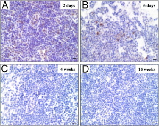
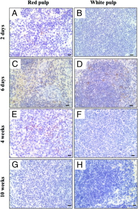
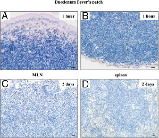
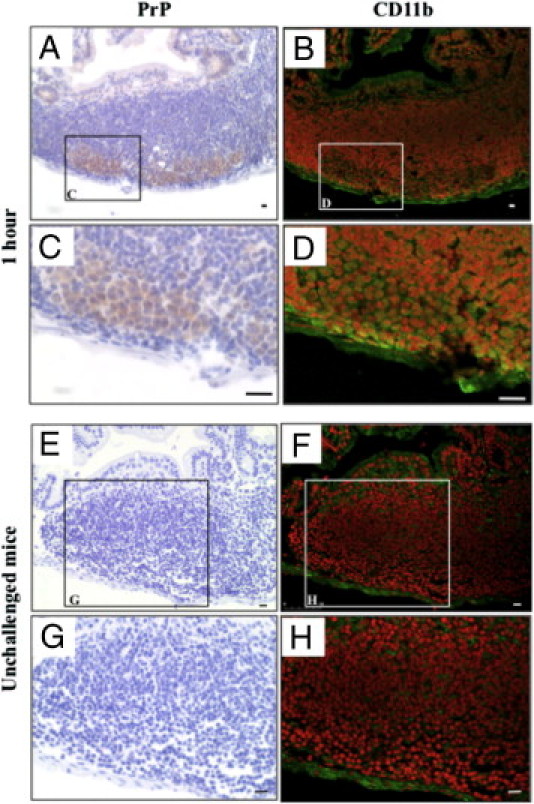
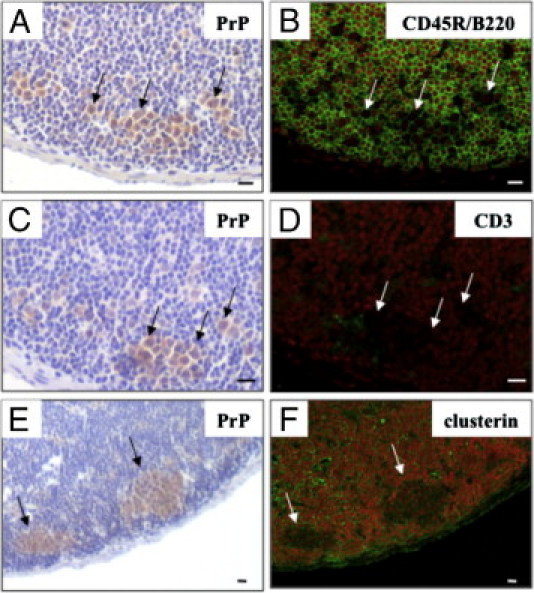
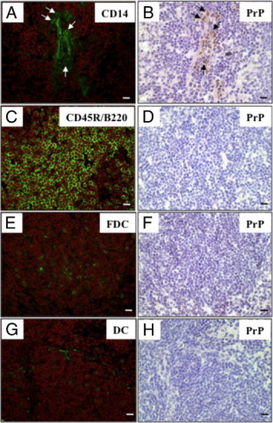
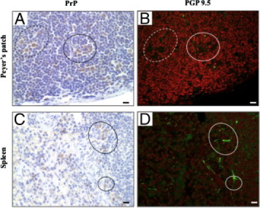
Transmissible spongiform encephalopathies are fatal neurodegenerative diseases. Infection by the oral route is assumed to be important, although its pathogenesis is not understood. Using prion protein (PrP) knockout mice, we investigated the sequence of events during the invasion of orally administered PrPs through the intestinal mucosa and the spread into lymphoid tissues and the peripheral nervous system. Orally administered PrPs were incorporated by intestinal epitheliocytes in the follicle-associated epithelium and villi within 1 hour. PrP-positive cells accumulated in the subfollicle region of Peyer's patches a few hours thereafter. PrP-positive cells spread toward the mesenteric lymph nodes and spleen after the accumulation of PrPs in the Peyer's patches. The number of PrP molecules in the mesenteric lymph nodes and spleen peaked at 2 days and 6 days after inoculation, respectively. The epitheliocytes in the follicle-associated epithelium incorporating PrPs were annexin V-positive microfold cells and PrP-positive cells in Peyer's patches and spleen were CD11b-positive and CD14-positive macrophages. Additionally, PrP-positive cells in Peyer's patches and spleen were detected in the vicinity of peripheral nerve fibers in the early stages of infection. These results indicate that orally delivered PrPs were incorporated by microfold cells promptly after challenge and that macrophages might act as a transporter of incorporated PrPs from the Peyer's patches to other lymphoid tissues and the peripheral nervous system.


Figures

Figure 1
Figure 1
Distribution of orally inoculated prion proteins (PrPs) in the Peyer's patches of the duodenum. Cellular PrP knockout mice were orally challenged with 200 μL of 10% Fukuoka-1 strain Creutzfeldt-Jakob disease brain homogenate. Small intestines were obtained at the indicated times after the challenge and subjected to immunostaining for PrPs. The paraffin sections of Peyer's patches were stained with anti-PrP antibody. Immunohistochemical photographs show the distribution of PrPs in the duodenum villi (A) at 1 hour and in Peyer's patch at 1 hour [B: lower magnification, C: subepithelial dome (SED), D: subfollicle], 4 hours (E: SED, F: subfollicle), and 8 hours (G: SED, H: subfollicle) after the challenge. Arrows indicate epitheliocytes including PrP-positive particle in the villi (A) and follicle-associated epithelium (FAE) (C) at 1 hour after the challenge. Scale bars = 10 μm.
Figure 2
Figure 2
Prion protein (PrP)-positive cells in the mesenteric lymph nodes (MLN). Then after oral challenge with 10% Fukuoka-1 strain Creutzfeldt-Jakob disease brain homogenate, MLN were obtained from cellular PrP (PrPc) knockout mice at 2 days (A), 6 days (B), 4 weeks (C), and 10 weeks (D). The paraffin sections of MLN were stained with anti-PrP antibody. Scale bars = 10 μm.
Figure 3
Figure 3
Prion protein (PrP)-positive cells in the spleen. Spleens were obtained from cellular PrP knockout mice at 2 days (A, B), 6 days (C, D), 4 weeks (E, F), and 10 weeks (G, H) after an oral challenge with 10% Fukuoka-1 strain Creutzfeldt-Jakob disease brain homogenate. The paraffin-embedded spleens were stained with anti-PrP antibody. The photographs show the area of red pulp (left column) and white pulp (right column). Scale bars = 10 μm.
Figure 4
Figure 4
Oral inoculation of normal brain homogenate. Cellular prion protein (PrPc) knockout mice were orally challenged with 200 μL of 10% normal brain homogenate. The small intestines, mesenteric lymph nodes (MLN), and spleen were obtained at the indicated times after the challenge and were subjected to immunostaining for prion proteins (PrPs). Immunohistochemical photographs show subepithelial dome (A) and subfollicle (B) regions of the duodenum Peyer's patch at 1 hour, MLN (C) at 2 days, and spleen (D) at 2 days after the challenge. Scale bars = 10 μm.
Figure 5
Figure 5
Prion proteins (PrP)-positive cells expressing annexin V in the follicle-associated epithelium. The same paraffin section of duodenum at 1 hour after the oral challenge was immunohistochemically re-stained, initially using anti-PrP antibody (A: with boxed areas magnified in C and E) and then with an anti-annexin V antibody (B: with boxed areas magnified in D and F). Arrows and arrowheads indicate the same PrP-positive cells. Scale bars = 10 μm.
Figure 6
Figure 6
Accumulation of prion protein (PrP)-positive macrophages in the subfollicle region of Peyer's patches. The small intestines were obtained from mice at 1 hour after the oral challenge (AD) or from unchallenged mice (EH). The same paraffin section of the Peyer's patches of the duodenum were immunohistochemically re-stained, initially using an anti-PrP antibody (left column) and then with an anti-CD11b antibody (right column). The boxed areas in A, B, E, and F are enlarged in C, D, G, and H, respectively. Scale bars = 10 μm.
Figure 7
Figure 7
Other cell types of the Peyer's patches of the duodenum did not stain for prion proteins (PrPs). The same paraffin section of the duodenum Peyer's patches at 1 hour after the oral challenge were immunohistochemically re-stained, initially using an anti-PrP antibody (A, C, E) and then with anti-CD45R/B220 (B), anti-CD3 (D), or anti-clusterin (F) antibodies. Arrows indicate the same PrP-positive cells. Scale bars = 10 μm.
Figure 8
Figure 8
Immunohistochemical re-staining of various cell surface markers and prion proteins (PrPs) in the spleen. The same frozen sections of spleen at 2 days after the oral challenge were immunohistochemically re-stained, initially using an anti-PrP antibody (B, D, F, H) and then with anti-CD14 (A), anti-CD45R/B220 (C), anti-follicular dendritic cells (FDC) (E) and anti-dendritic cell (DC) (G) antibodies. Arrows indicate the same PrP-positive cells. Scale bars = 10 μm.
Figure 9
Figure 9
Immunohistochemical re-staining of prion proteins (PrPs) and nerve fibers in Peyer's patches and the spleen. The samples of Peyer's patches and spleen were obtained from mice at 4 hours and 2 days after an oral challenge, respectively. The same paraffin sections were immunohistochemically re-stained initially using an anti-PrP antibody (A,C) and then with an anti-protein gene product 9.5 antibody (B, D). Dotted and solid circles in the left column correspond to those in the right column. Scale bars = 10 μm.

Similar articles

Cited by

References

    1. Aguzzi A. Prion diseases of humans and farm animals: epidemiology, genetics and pathogenesis. J Neurochem. 2006;97:1726–1739. - PubMed
    1. Masters C.L., Richardson E.P., Jr Subacute spongiform encephalopathy (Creutzfeldt-Jakob Disease): The nature and progression of spongiform change. Brain. 1978;101:333–344. - PubMed
    1. van Everbroeck B., Dobbeleir I., De Waele M., De Leenheir E., Lubke U., Martin J.J., Cras P. Extracellular protein deposition correlates with glial activation and oxidative stress in Creutzfeldt-Jakob and Alzheimer's disease. Acta Neuropathol. 2004;108:194–200. - PubMed
    1. van Keulen L.J., Vromans M.E., Dolstra C.H., Bossers A., van Zijderveld F.G. Pathogenesis of bovine spongiform encephalopathy in sheep. Arch Virol. 2008;153:445–453. - PMC - PubMed
    1. Bolton D.C., McKinley M.P., Prusiner S.B. Identification of a protein that purifies with the scrapie prion. Science. 1982;218:1309–1311. - PubMed

Publication types