. 2011 Sep;179(3):1483-93.
doi: 10.1016/j.ajpath.2011.05.060. Epub 2011 Jul 18.

Pleural mesothelioma instigates tumor-associated fibroblasts to promote progression via a malignant cytokine network

Affiliations

Pleural mesothelioma instigates tumor-associated fibroblasts to promote progression via a malignant cytokine network

Qi Li et al. Am J Pathol. 2011 Sep.

Abstract

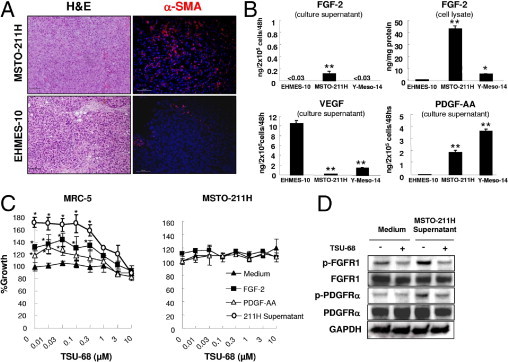
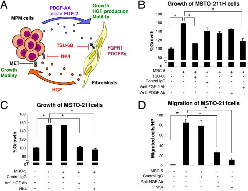
The tumor microenvironment is crucial to the progression of various malignancies. Malignant pleural mesothelioma (MPM), which originates from the pleura, grows aggressively in the thoracic cavity. Here we describe an orthotopic implantation SCID mouse model of MPM and demonstrate that α-SMA-positive fibroblast-like cells accumulate in the tumors produced by the human MPM cell lines MSTO-211H and Y-Meso-14. We assessed the interaction between MPM cells and their microenvironments, focusing on tumor-associated fibroblasts. MSTO-211H and Y-Meso-14 cells produced fibroblast growth factor-2 (FGF-2) and/or platelet-derived growth factor-AA (PDGF-AA); they also enhanced growth, migration, and production of hepatocyte growth factor (HGF) by human lung fibroblast MRC-5 cells. MRC-5 cells stimulated HGF-mediated growth and migration of MSTO-211H and Y-Meso-14 cells in an in vitro coculture system. In the orthotopic model, tumor formation by MSTO-211H and Y-Meso-14 cells was significantly inhibited by TSU-68, an inhibitor of FGF, VEGF, and PDGF receptors; imatinib, an inhibitor of PDGF receptors; and NK4, an antagonist of HGF. Histological analyses of clinical specimens from 51 MPM patients revealed considerable tumor-associated fibroblasts infiltration and expression of HGF, together with FGF-2 or PDGF-AA, in tumors. These findings indicate that MPM instigates tumor-associated fibroblasts, promoting tumor progression via a malignant cytokine network. Regulation of this cytokine network may be therapeutically useful for controlling MPM.


Figures

Figure 1
Figure 1
MSTO-211H cell-derived FGF-2 and PDGF-AA promote growth of fibroblasts. A: Thoracic tumors produced by MPM cells in SCID mice were stained with H&E and by immunofluorescence for α-SMA (red) and DAPI (blue). Scale bars = 100 μm. B: ELISA assays of FGF-2, VEGF, and PDGF-AA in the culture supernatant of MPM cells and of FGF-2 in cell lysates. *P < 0.05, **P < 0.01 versus EHMES-10. C: MRC-5 or MSTO-211H cells were cultured in the presence of FGF-2 (20 ng/mL), PDGF-AA (20 ng/mL), or the culture supernatant of MSTO-211H cells (2 × 10⁵/2 mL/48 hours), with or without TSU-68. After 72 hours, the growth of MRC-5 or MSTO-211H cells was determined by MTT assay. *P < 0.01 versus medium alone. D: TSU-68 abrogates the phosphorylation of FGFR1 and PDGFRα induced by the supernatant of MSTO-211H cells. MRC-5 cells were incubated with or without TSU-68 (10 μmol/L) in the presence or absence of the supernatant of MSTO-211H cells for 15 minutes. The cells were lysed and proteins were detected by immunoblotting.
Figure 2
Figure 2
HGF derived from fibroblasts enhances the growth and motility of MPM cells. A: HGF production by MPM (EHMES-10, MSTO-211H), fibroblast (MRC-5 and IMR-90), Primary cultured patient fibroblast (PF), and endothelial (HMVEC) cell lines. Cells (2 × 10⁵/2 mL) were incubated in medium for 8 hours, the culture supernatants were harvested, and HGF concentrations were determined by ELISA. B: FGF-2 and PDGF-AA enhance HGF production by fibroblasts. MRC-5 cells (2 × 10⁵/2 mL) were cultured for 48 hours in the presence of FGF-2 (20 ng/mL) or PDGF-AA (20 ng/mL), and HGF concentrations in culture supernatants were determined by ELISA. *P < 0.05; **P < 0.01 versus medium alone. C: FGF-2 and PDGF-AA increase HGF mRNA expression in fibroblasts. HGF mRNA expression by MRC-5 cells pretreated with FGF-2 (20 ng/mL) or PDGF-AA (20 ng/mL) for 48 hours was determined by real-time RT-PCR. *P < 0.05; **P < 0.01 versus medium alone. D: HGF increases MPM cell growth. MSTO-211H cells were cultured with 20 ng/mL of HGF, FGF-2, or PDGF-AA in the presence or absence of NK4 (300 nmol/L) for 72 hours, and cell growth was determined by MTT assay. *P < 0.05, **P < 0.01 versus HGF. E: NK4 abrogates MET phosphorylation induced by fibroblast-derived HGF in MPM cells. MSTO-211H cells were treated with or without NK4 (300 nmol/L) in the presence or absence of HGF (20 ng/mL) or the supernatant of MRC-5 cells (106/mL/48 hours) for 30 minutes. The cells were lysed and proteins were detected by immunoblotting. The lower band (p145 MET) corresponds to the mature form of MET; the upper band (p170 MET) corresponds to the immature MET precursor. F: HGF induces the migration of MSTO-211H cells. The assay was performed in triplicate as described under Materials and Methods. *P < 0.05, **P < 0.01 versus medium alone.
Figure 3
Figure 3
Simultaneous blockade of FGFR1 and PDGFRs inhibits the growth of MPM cells in a coculture system. A: Schema of the malignant cytokine network between MPM cells and stromal tumor-associated fibroblasts (TAFs). B: TSU-68 circumvents the growth of MPM cells induced by fibroblasts in the coculture system. MSTO-211H cells were cocultured with MRC-5 cells with or without TSU-68 (10 μmol/L), control IgG, anti-FGF-2 antibody, and/or anti-PDGF antibody (1 μg/mL) for 72 hours. MPM cell growth was determined using a Cell Counting Kit-8. C: Inhibition of HGF blocks the growth of MPM cells induced by fibroblasts in a coculture system. MSTO-211H cells were cocultured with MRC-5 cells with control IgG, anti-HGF neutralizing antibody (1 μg/mL), or NK4 (300 nmol/L) for 72 hours, and MPM cell growth was determined. D: Blockade of HGF prevents MSTO-211H cell migration induced by MRC-5 cells. The assay was performed in triplicate as described under Materials and Methods. *P < 0.01.
Figure 4
Figure 4
Malignant cytokine network between MPM cells and patient fibroblasts or mouse fibroblasts. A: Blockade of HGF prevents MSTO-211H or Y-Meso-14 cell migration induced by primary cultured patient fibroblasts (PF). The assay was performed in triplicate as described under Materials and Methods. *P < 0.05, **P < 0.01 versus PF. B and C: TSU-68, NK4, and anti-HGF antibody blocked the growth of MSTO-211H cells induced by PF in the coculture system. MSTO-211H (B) or Y-Meso-14 (C) cells were cocultured with PF cells with or without TSU-68 (10 μmol/L), control IgG, NK4 (300 nmol/L), anti-HGF antibody (1 μg/mL), anti-FGF-2 antibody, and/or anti-PDGF antibody (1 μg/mL) for 72 hours. MPM cell growth was determined using a Cell Counting Kit-8. *P < 0.05, **P < 0.01 versus PF. D: PDGF-AA (20 ng/mL), but not FGF-2 (20 ng/mL), enhances the growth of the mouse fibroblast 3T3-Swiss cell line, as determined by MTT assays. *P < 0.05. E: NK4 abrogates MET phosphorylation induced by mouse fibroblast-derived HGF in MPM cells. MSTO-211H cells were treated with or without NK4 (300 nmol/L) in the presence or absence of mouse HGF (100 ng/mL) or the supernatant of 3T3-Swiss cells (106/mL/48 hours) for 30 minutes. The cells were lysed and proteins were detected by immunoblotting. F: NK4 decreases the migration of MPM cells induced by mouse fibroblasts. Migration of MSTO-211H cells and Y-Meso-14 cells in the presence or absence of NK4 (300 nmol/L) cocultured with 3T3-Swiss cells was assayed as described under Materials and Methods. *P < 0.05, **P < 0.01.
Figure 5
Figure 5
TSU-68, imatinib, and NK4 inhibit the production of thoracic tumors by MPM cells in an orthotopically implanted SCID mouse model. A: Macroscopic appearance of thoracic tumors caused by MSTO-211H or Y-Meso-14 cells treated with TSU-68, imatinib, or NK4. The thoracic tumors were evaluated as described under Materials and Methods. B: Histological analysis of the same tumors. The thoracic tumors were analyzed by immunohistochemistry (type I collagen and BrdU) and immunofluorescence (α-SMA and double staining of BrdU and type I collagen). In double staining, red fluorescence indicates BrdU and green fluorescence indicates type I collagen; arrows mark double-positive cells (proliferating fibroblasts). Scale bars = 50 μm. The corresponding graphs show the mean number of positive cells per high-power field (HPF), from five independent areas. *P < 0.01 versus control. Negative controls without primary antibodies did not show discernible staining for α-SMA, BrdU, or type I collagen (data not shown).

References

    1. Carbone M., Kratzke R.A., Testa J.R. The pathogenesis of mesothelioma. Semin Oncol. 2002;29:2–17. - PubMed
    1. Robinson B.W., Lake R.A. Advances in malignant mesothelioma. N Engl J Med. 2005;353:1591–1603. - PubMed
    1. Aisner J. Current approach to malignant mesothelioma of the pleura. Chest. 1995;107:332S–344S. - PubMed
    1. Vogelzang N.J., Rusthoven J.J., Symanowski J., Denham C., Kaukel E., Ruffie P., Gatzemeier U., Boyer M., Emri S., Manegold C., Niyikiza C., Paoletti P. Phase III study of pemetrexed in combination with cisplatin versus cisplatin alone in patients with malignant pleural mesothelioma. J Clin Oncol. 2003;21:2636–2644. - PubMed
    1. Bhowmick N.A., Neilson E.G., Moses H.L. Stromal fibroblasts in cancer initiation and progression. Nature. 2004;432:332–337. - PMC - PubMed

Publication types

MeSH terms

Substances