Comparative Study
. 2012 Jul;176(1):321-8.
doi: 10.1016/j.jss.2011.05.041. Epub 2011 Jun 22.

Local and systemic alterations in signal transducers and activators of transcription (STAT) associated with human abdominal aortic aneurysms

Affiliations
Comparative Study

Local and systemic alterations in signal transducers and activators of transcription (STAT) associated with human abdominal aortic aneurysms

Mingfang Liao et al. J Surg Res. 2012 Jul.

Abstract

Background: Signal transducers and activators of transcription (STAT) proteins are transcription factors that, when activated by phosphorylation, regulate gene expression and cellular activity. The aim of this study was to evaluate the local and systemic expression and activation of STAT proteins associated with abdominal aortic aneurysms (AAA).

Methods: Expression and activation of STAT proteins were assessed in aortic wall samples obtained from patients undergoing repair of AAA (n = 9) and from non-aneurysmal (NA) donors (n = 17). Aortic samples were evaluated for mRNA and protein expression for STAT1, 2, 3, 4, 5a, and 5b using RT-PCR and immunoblot (WB) assays and normalized to ß-actin (expressed as arbitrary units). STAT activation was assessed with WB assays using phosphorylated (p)-STAT-specific antibodies. Alterations in STAT activation were calculated by normalizing pSTAT proteins to corresponding total STAT levels. Immunohistochemistry was performed on AAA and NA samples using the total and pSTAT antibodies. Systemic alterations in STAT activation were assessed by evaluating circulating leukocytes for the presence of pSTAT from patients with AAA (AAA, n = 8), repaired aneurysm (RA, n = 8), or age/gender matched controls with no AAA (CT, n = 8). Flow cytometry was performed to assess for circulating levels of STAT1 (pY701), STAT3 (pY705), and STAT5a (pY694) in monocytes, granulocytes, and lymphocytes. Assessments were made at baseline and in response to in vitro stimulation with IFN-γ (50 ng/mL) or IL-6 (100 ng/mL). Results were analyzed using Student's t-test and are expressed as mean ± SEM.

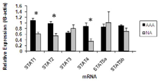
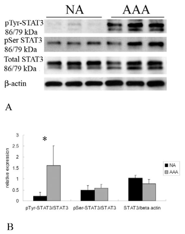
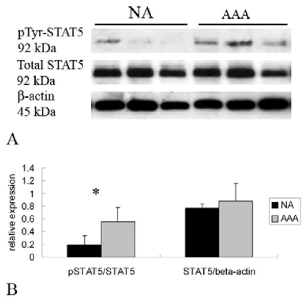
Results: In AAA tissue compared with NA, STAT-1 (1.08 ± 0.09 versus 0.62 ± 0.07), -2 (0.98 ± 0.07 versus 0.55 ± 0.08), and -4 (0.89 ± 0.12 versus 0.35 ± 0.11) mRNA levels were elevated (P < 0.01, all). Corresponding increases in STAT protein were only observed for STAT1 (2.77 ± 0.93 versus 0.93 ± 0.08, P < 0.05). Increases in activation were observed in AAA compared with NA in pSTAT2 (0.77 ± 0.1 versus 0.1 ± 0.02, P < 0.01), pSTAT3 (1.6 ± 0.3 versus 0.2 ± 0.06, P < 0.02) and pSTAT5 (0.57 ± 0.03 versus 0.2 ± 0.03, P < 0.05) levels. Phosphorylated STAT1, 2, 3, and 5 were observed in inflammatory cells invading the AAA adventitia. In addition, STAT3 was observed in the media of AAA and NA, but pSTAT3 was only observed in the media of AAA. There were no differences in baseline levels of pSTAT-positive circulating leukocytes. IFN-γ stimulation decreased STAT-5a (pY694)-positive CT lymphocytes to 40% ± 13% of baseline, but had no effect on AAA or RA lymphocytes (116% ± 35%, 102% ± 19%, respectively; P = 0.01). STAT-5a (pY694)-positive CT granulocytes also decreased to 62% ± 18% of baseline compared with AAA or RA granulocytes (122% ± 25%, 126% ± 17%, respectively; P = 0.01). Alterations in STAT1 (pY701) and STAT3 (pY705) were not observed in leukocytes following cytokine stimulation.

Conclusions: STAT proteins are important regulators of transcriptional activity and have been linked to cardiovascular disease. The present data suggest that altered levels of phosphorylated STATs are associated with AAA. Understanding their role may provide further insight into the mechanisms of AAA formation and allow for the development of medical treatment options.


Figures

Figure 1
Figure 1
Graphic representation of aortic STAT mRNA obtained from AAA (black bars) or NA (gray bars). STAT mRNA expression was assessed by real time RT-PCR and normalized to β-actin and is expressed in arbitrary units (AU). STAT1, 2, and 4 mRNA were significantly elevated in samples obtained from AAA compared to NA. * P<0.05.
Figure 2
Figure 2
Graphic representation of aortic total STAT protein obtained from AAA (black bars) or NA (gray bars). STAT protein expression was assessed from aortic homogenates using immunoblot assay. Samples were normalized to β-actin and are expressed in arbitrary units. Only STAT1 levels were significantly elevated in AAA samples compared to NA controls (*P<0.01).
Figure 3
Figure 3
(A) Representative immunoblots from aortic homogenates obtained from those without aneurysms (NA) or with aneurysms (AAA). Samples were blotted against p(Y690)STAT-2, total STAT-2 and β-actin. Assessment for STAT2 activation was assessed by analyzing the pSTAT2 versus total STAT2. (B) Graphic representation of STAT2 activation obtained from NA and AAA samples. STAT2 underwent significantly more phosphorylation in samples from AAA compared to those from NA (*P<0.01). There were no significant changes in total STAT2 levels.
Figure 4
Figure 4
(A) Representative immunoblots from aortic homogenates obtained from those without aneurysms (NA) or with aneurysms (AAA). Samples were blotted with antibodies against p(Y705)STAT-3, p(S727)STAT-3, total STAT3, and β-actin. Assessment for STAT3 activation was assessed by analyzing the different p-STAT-3 versus total STAT-3 (B) Graphic representation of STAT3 activation obtained from NA and AAA samples. STAT3 underwent significantly more activation by tyrosine phosphorylation in AAA compared to NA controls (*P<0.01). There were no significant differences in serine phosphorylation or in total STAT3 levels.
Figure 5
Figure 5
(A) Representative immunoblots from aortic homogenates obtained from those without aneurysms (NA) or with aneurysms (AAA). Samples were blotted with antibodies against p(Y694)STAT5, total STAT5, and β-actin. (B) Assessment of STAT5 activation was determined by assessing the ratio of p-STAT5 to total STAT5. While there was no difference in total STAT5 protein from AAA (gray bar) compared to NA (black bar), there was a significant increase in STAT5 activation demonstrated by the significant increase in the ratio of p-STAT5/STAT5 (*P<0.01).
Figure 6
Figure 6
Representative samples from aortic tissue obtained from non-aneurysmal aorta (A and C) and aneurysmal aorta (B and D). Immunohistochemical analysis failed to show any staining for STAT1 in non-aneurysmal tissue (A (40x) and C (100x)), but was present (arrow) in the adventitial layer in samples from aortic aneurysms (B, 40x). This appeared to localize to inflammatory cells (arrows) within the adventitial layer (D, 100x). Corresponding cells in this layer also stained positive for CD68 (E, 100x) suggesting they are of a monocyte/macrophage lineage.
Figure 7
Figure 7
Representative sample from aortic tissue from a patient with (B (40x), C (100X) and D (100x)) and without (A (40X)) an AAA. Immunohistochemical analysis with antibodies directed against STAT5 failed to demonstrate any positive staining in the normal aorta (A), but there was positive staining for both STAT5 (B) and pSTAT5 (C) in inflammatory cells infiltrating the adventitia of the aneurysm. Similarly, staining with antibodies to pSTAT2 showed positive cells in the adventitial inflammatory cells (D).
Figure 8
Figure 8
Representative samples from aortic tissue obtained from non-aneurysmal aorta (A and B (100x)) and aneurysmal tissue (C, D, E (100x)). Immunohistochemical analysis demonstrates that STAT3 staining (brown cells) occurs in both non-aneurysmal (A) and aneurysmal (C) tissue in the medial layer in smooth muscle cells. pSTAT3 staining, however, only occurred in aneurysmal tissue (arrow) (D) and not non-aneurysmal aorta (B). STAT3 (not shown) and pSTAT3 also stained positive in the inflammatory cells within the adventitia of aneurysms (E).

Similar articles

Cited by

References

    1. Saratzis A, Kitas GD, Saratzis N, Melas N. Can statins suppress the development of abdominal aortic aneurysms? A review of the current evidence. Angiology. 2010;61:137–44. - PubMed
    1. Shimizu K, Mitchell RN, Libby P. Inflammation and cellular immune responses in abdominal aortic aneurysms. Arterioscler Thromb Vasc Biol. 2006;26(5):987–94. - PubMed
    1. Shimizu K, Shichiri M, Libby P, Lee R, Mitchell R. Th2-predominant inflammation and blockade of IFN gamma signaling induce aneurysm in allografted aortas. J Clin Invest. 2004;114:168–71. - PMC - PubMed
    1. King V, Yin A, Kristo F, Anderson T, Ahluwalia N, Hardy G, et al. Interferon-gamma and the interferon-inducible chemokine CXCL10 protect against aneurysm formation and rupture. Circulation. 2009;119:426–35. - PMC - PubMed
    1. Ocana E, Perez-Requena J, Bohorquez J, Brieva J, Rodriguez C. Chemokine receptor expression on infiltrating lymphocytes from abdominal aortic aneurysms: role of CXCR4-CXCL12 in lymphoid recruitment. Atherosclerosis. 2008;200:264–70. - PubMed

Publication types