Review
. 2012 Mar;30(3):138-46.
doi: 10.1016/j.tibtech.2011.06.013. Epub 2011 Jul 20.

Self-folding devices and materials for biomedical applications

Affiliations
Review

Self-folding devices and materials for biomedical applications

Christina L Randall et al. Trends Biotechnol. 2012 Mar.

Abstract

Because the native cellular environment is 3D, there is a need to extend planar, micro- and nanostructured biomedical devices to the third dimension. Self-folding methods can extend the precision of planar lithographic patterning into the third dimension and create reconfigurable structures that fold or unfold in response to specific environmental cues. Here, we review the use of hinge-based self-folding methods in the creation of functional 3D biomedical devices including precisely patterned nano- to centimeter scale polyhedral containers, scaffolds for cell culture and reconfigurable surgical tools such as grippers that respond autonomously to specific chemicals.


Figures

Figure 1
Figure 1. Schematic and versatility of the self-folding method for hollow polyhedral containers
(a) Schematic showing 2D to 3D self-folding of a dodecahedron using surface tension based self-folding. The 2D template or net is patterned with hinges between panels (folding hinges) and also at the edges (locking hinges). (b) Schematics showing functionality of the two hinges, (b1) folding hinges provide a torque to rotate panels and (b2) locking hinges self-align and fuse panels at non-folding edges. Folding and locking are both driven by physical forces associated with the minimization of surface area of the molten hinge materials. Adapted, with permission, Ref. [33] ©IOP Publishing Ltd. (c) Video capture sequence (over 15 s) showing self-folding of a 1 mm sized, six-windowed polymeric container (with SU-8 panels and biodegradable polycaprolactone (PCL) hinges) on heating to 60°C. (d). Fluorescence image of a group of 1 mm sized SU8/PCL containers. (e) Fluorescence z-plane stack image of live, calcein stained fibroblast cells encapsulated within an optically transparent SU8/PCL container. Reprinted, with permission, Ref. [26] ©Springer. (f) SEM image of a self-folded dodecahedral shaped hollow metallic container featuring anisotropic surface patterning of slits. (g) Optical image of numerous 100 µm, 200 µm and 500 µm (panel edge length) dodecahedra, highlighting that many containers can be fabricated en masse. Reprinted, with permission, Ref. [33] ©IOP Publishing Ltd. (h–i) SEM images of electron-beam patterned 2D templates and self-folded 100 nm scale cubic particles. It should be noted that the self-folding process is parallel even at the nanoscale and the particles have the letters JHU patterned with line widths as small as 15 nm. These particles have been created with both metallic (nickel) and ceramic (alumina) panel composition. Reprinted, with permission, Ref. [25] ©American Chemical Society 2009. The scale bars: (a–e) 500 µm and (f–d) 250 nm long.
Figure 2
Figure 2. Versatile design of 3D microwell arrays composed of self-folding devices for enhanced diffusion in biomedical applications
(a). Conceptual schematic of a conventional 2D microwell array (top) and our 3D microwell array (bottom). The important difference is that although both are 3D recesses, microfabricated 2D microwell arrays feature well defined porosity only along one surface. (b–c) Optical images of ordered 3D microwell arrays composed of Au-coated containers on both (b) flat polyurethane coated silicon and (c) curved polymeric surfaces. The number “3” and letter “D” are spelt out to highlight versatility in the spacing and positioning offered by this technique. All scale bars are 500 µm long. (d) Insulin response profiles to a glucose stimulation from one, three and five porous-faced microwell arrays after seven days. Data are plotted as the average ± the standard deviation (sample size n = 5). (e) A graph showing the four hour (steady-state) insulin concentration measured in response to a glucose stimulation for β-TC-6 cells encapsulated within 2D (one porous-faced), three porous-faced and 3D (five porous-faced) microwell arrays. The average and the standard deviation obtained on days 1 (number of samples, n, = 5), 7 (n = 5), 14 (n = 3) and 28 (n = 3) are plotted. The 3D microwell arrays produced significantly greater stimulated insulin at longer times. Reprinted, with permission, Ref. [38] ©The Royal Society of Chemistry.
Figure 3
Figure 3. Using containers with precisely patterned wall porosity to direct the chemotactic self-organization of E. coli in the shape of a helix
(a–b). A conceptual schematic of the desired chemotactic self organization in a spiral. At the start of the experiment, a) the chemoattractant is confined to the Au-coated container and green fluorescent protein (GFP) expressing E. coli cells are distributed uniformly throughout the medium. b) E. coli cells self-organize in a helical pattern based on the underlying chemical pattern once the chemoattractant (L-serine, yellow) is allowed to diffuse out of the container. (c–d) Experimental realization of the concept. Time-lapse images of green fluorescent E. coli as they self-organized in a helical pattern around a container. The scale bar is 500 µm long. Adapted and reprinted with permission, Ref. [42] ©Wiley-VCH Verlag GmbH & Co. KgaA.
Figure 4
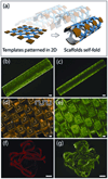
Figure 4. Self-folding scaffolds in anatomically relevant geometries
(a) Schematic of self-folding of planar, micropatterned templates from 2D to 3D geometries on release from the substrate. (b–c) Fluorescent image of a calcein stained cell culture grown on cylindrical scaffolds of two different diameters. The cylinders consist of a 15 × 15 array of 160 µm square panels spaced 80 µm apart. (d–e) Optical images of a bi-directionally folded scaffold and fluorescently labeled fibroblasts cultured on it. Schematic overlay illustrates the undulatory cross-section of the scaffold. (f–g) Optical images of a self-folding spiral-like ribbon and calcein stained fibroblasts cultured on it. The scale bars are 160 µm long. Adapted and reprinted with permission, Ref. [56] ©Elsevier, 2010.
Figure 5
Figure 5. Self-folding miniaturized tools for surgery
(a–e) Optical microscopy sequence showing capture and retrieval of neutral red-stained cells from a cell culture mass at the end of a tube; the scale bar is 1 mm long. (f) Fluorescent micrograph of viable (green) L929 cells captured by using a biochemical trigger to actuate the gripper; the scale bar is 100 µm long. (g) Optical image of a microgripper with a tissue sample retrieved from a bovine bladder; the scale bar is 100 µm long. Adapted and reprinted with permission, Ref. [69]. (h, i) Body temperature activated microgrippers in intrahepatic porcine bile ducts, ex-vivo, The scale bar is 1 mm long.
Figure 6
Figure 6. Surgical tools that close and open when exposed to enzymes
(a–c) Optical images of a microgripper patterned with gelatin (a polypeptide) and carboxymethylcellulose (CMC, a polysaccharide) triggers in three different states: open (as fabricated), closed (on exposure to cellulase), and re-opened (on exposure to proteases such as collagenase). The red arrow indicates the set of hinges that cause re-opening. The scale bars are 200 µm long. (d–f) Schematic conceptual representations of the grippers in the three corresponding states. (d) The gripper is kept flat by the biopolymer layers. (e) When one biopolymer is selectively degraded by a specific enzyme class (enzyme 1) the gripper closes. (f) Subsequently, a second set of hinges, which are insensitive to enzyme 1 are actuated by another enzyme class (enzyme 2) and the gripper re-opens (g) Plots of the kinetics of gripper closing on exposure to different enzymes indicating cross-selectivity of over two orders of magnitude. Adapted and reprinted with permission, Ref. [70] ©American Chemical Society, 2009.

References

    1. Madou MJ. Fundamentals of microfabrication: the science of miniaturization. 2nd ed. CRC Press; 2002.
    1. Charnley M, et al. Integration column: microwell arrays for mammalian cell culture. Integr Biol. 2009;1:625–634. - PubMed
    1. Desai TA, et al. Microfabricated immunoisolating biocapsules. Biotechnol Bioeng. 1998;57:118–120. - PubMed
    1. Rappaport C. Review—Progress in concept and practice of growing anchorage-dependent mammalian cells in three dimensions. In Vitro Cell Dev Biol Anim. 2003;39:187–192. - PubMed
    1. Santini JT, et al. Microchips as controlled drug-delivery devices. Angew Chem Int Ed Engl. 2000;39:2396–2407. - PubMed

Publication types

Substances