. 2011 Jul 15;333(6040):348-53.
doi: 10.1126/science.1205822.

Precise manipulation of chromosomes in vivo enables genome-wide codon replacement

Affiliations

Precise manipulation of chromosomes in vivo enables genome-wide codon replacement

Farren J Isaacs et al. Science. .

Abstract

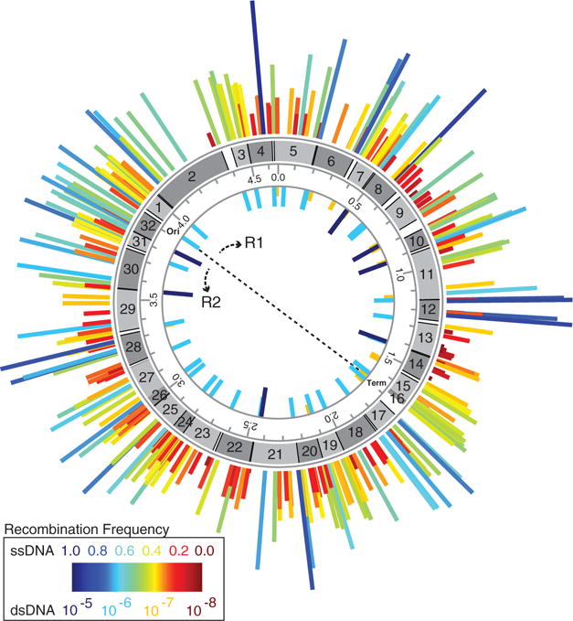
We present genome engineering technologies that are capable of fundamentally reengineering genomes from the nucleotide to the megabase scale. We used multiplex automated genome engineering (MAGE) to site-specifically replace all 314 TAG stop codons with synonymous TAA codons in parallel across 32 Escherichia coli strains. This approach allowed us to measure individual recombination frequencies, confirm viability for each modification, and identify associated phenotypes. We developed hierarchical conjugative assembly genome engineering (CAGE) to merge these sets of codon modifications into genomes with 80 precise changes, which demonstrate that these synonymous codon substitutions can be combined into higher-order strains without synthetic lethal effects. Our methods treat the chromosome as both an editable and an evolvable template, permitting the exploration of vast genetic landscapes.


Figures

Fig. 1
Fig. 1
Strategy for reassigning all 314 TAG codons to TAA in E. coli. First, the genome was split into 32 regions each containing 10 TAG stop codons. In parallel, MAGE was used to execute all 10 TAG::TAA codon modifications in a single strain for each genomic region. These partially recoded strains were paired such that a targeted genomic region of one strain (donor) was strategically transferred into a second strain (recipient), permitting the hierarchical consolidation of modified genomic regions using CAGE (see Fig. 4A). This five-stage process transfers genomic fragments ranging in size from ~154 kb to ~2.3 Mb in a controlled manner until a single recoded strain is constructed that lacks the TAG stop codon throughout. Thus far, 28 of 31 conjugations have been completed, where the dotted arrows denote outstanding conjugation steps and dotted genomes represent half- and full-genome strains that have not yet been completed. Once all TAG codons have been converted to TAA, the prfA gene will be deleted to inactivate TAG translational termination.
Fig. 2
Fig. 2
Frequency map of oligo-mediated TAG::TAA codon replacements and genetic marker integrations across the E. coli genome at each replacement position. Circular map illustrates (from inner circle outward): (i) frequency of dsDNA selectable marker integrations; (ii) genome coordinates (in Mb): position of origin (Ori) and terminus (Term) and direction of the two replication forks (R1 and R2); (iii) location of the 32 targeted chromosomal segments; and (iv) frequency of TAG::TAA replacements across all TAG codons—after 18 MAGE cycles—denoted by height- and color-coded bars (scale bar indicates integration frequency).
Fig. 3
Fig. 3
Clonal rate and distribution of genome modifications after 18 cycles of MAGE. (A) Histogram of the frequencies of clones containing 1 to 10 conversions found among 1504 clones screened. A Poisson fit is shown (solid yellow line) for a subpopulation that excludes the zero-conversion group. (B) Distribution of modifications among the group of top clones (one for each of the 31 groups of 10 targeted modifications; one additional strain not shown had conversion at all four codon sites). (C) Distribution of the 314 90-mer oligos by their extent of total secondary sequence similarity to the E. coli genome. Inset box plot: Oligos with a mistarget score of more than 600 bp show, on average, a 32% decrease in allelic replacement (AR) frequency relative to oligos scoring less than 600 bp (25.6% versus 37.6%, P < 0.003).
Fig. 4
Fig. 4
Hierarchical CAGE methodology for controlled genome transfer. (A) Two pairs of strains illustrate the design and methodology of CAGE, with recoded genomic regions in red. Partially recoded strains are split into conjugation pairs. The donor strain (D) contains the following: oriT-kanamycin cassette (O, blue triangle); positive selection gene (Pn, n = 1, 2, or 3, black rectangle); and F plasmid (F+). The recipient strain (R) contains the following: positive-negative selection gene (PN) and Pn, flanking its recoded region. DNA transfer is initiated at O in the donor genome, ensuring transfer of the desired codon mutations and downstream Pn. After conjugation, a specific set of three simultaneous selections is applied to yield a recombinant strain that contains the recoded genomic fragment from the donor strain while retaining the other recoded region in the recipient genome. Placement of the PN marker downstream of the oriT sequence in the recipient genome ensures that the entire desired region of the donor genome is inherited in the recombinant strain. All conjugation factors are maintained episomally on F′, so only a ~2-kb oriT sequence must be inserted onto the genome to generate a highly controllable Hfr donor strain. Because there is no scar between the two recoded regions from the conjugation parents, only one recombination is required to insert the O (donor) or PN (recipient) directly into Pn for the next round of conjugation. This conjugation assembly-selection strategy is implemented in five stages to merge the genomes of 32 recoded strains into a single strain (see Fig. 1). (B) Genome transfer frequency as a function of the distance from O. Plots of two conjugations of genome segments illustrating the transfer of ~120 kb with positive and negative selection (blue) and ~360 kb with only positive selection (green) to assemble recoded genomic DNA from donor and recipient genomes. (C and D) MASC-PCR images of TAA alleles transferred under positive selection alone (C) and positive and positive-negative selection (D). Yellow arrows indicate the genomic point of transfer, which illustrate the inheritance of the donor TAA alleles in the conjugated strain.

Similar articles

Cited by

References

    1. Ambrogelly A, Palioura S, Söll D. Nat Chem Biol. 2007;3:29. - PubMed
    1. Knight RD, Freeland SJ, Landweber LF. Nat Rev Genet. 2001;2:49. - PubMed
    1. Young TS, Schultz PG. J Biol Chem. 2010;285:11039. - PMC - PubMed
    1. Neumann H, Wang K, Davis L, Garcia-Alai M. J W Chin, Nature. 2010;464:441. - PubMed
    1. Mukai T, et al. Nucleic Acids Res. 2010;38:8188. - PMC - PubMed

Publication types

Substances