. 2011 Sep 16;333(6049):1633-7.
doi: 10.1126/science.1207227. Epub 2011 Jul 14.

Sequence and structural convergence of broad and potent HIV antibodies that mimic CD4 binding

Affiliations

Sequence and structural convergence of broad and potent HIV antibodies that mimic CD4 binding

Johannes F Scheid et al. Science. .

Abstract

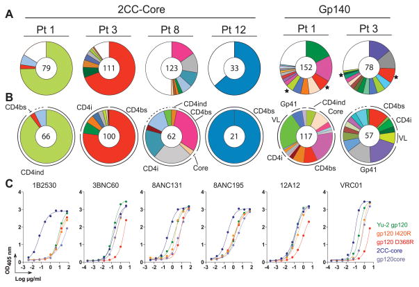
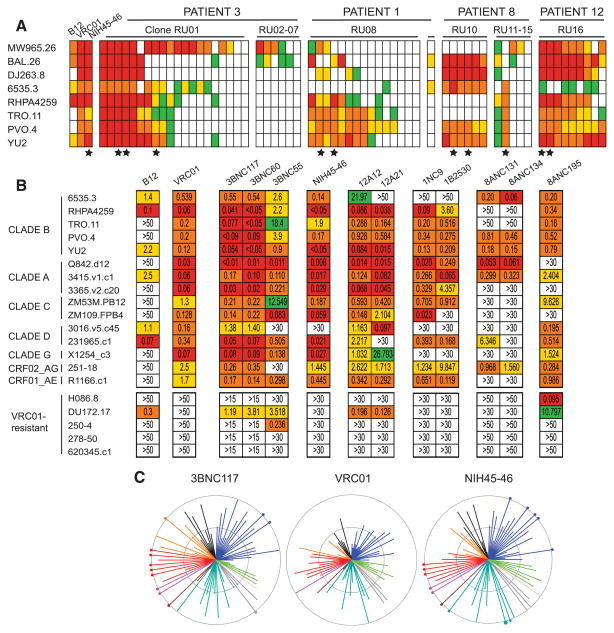
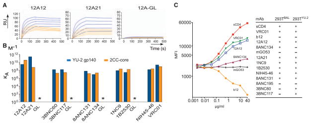
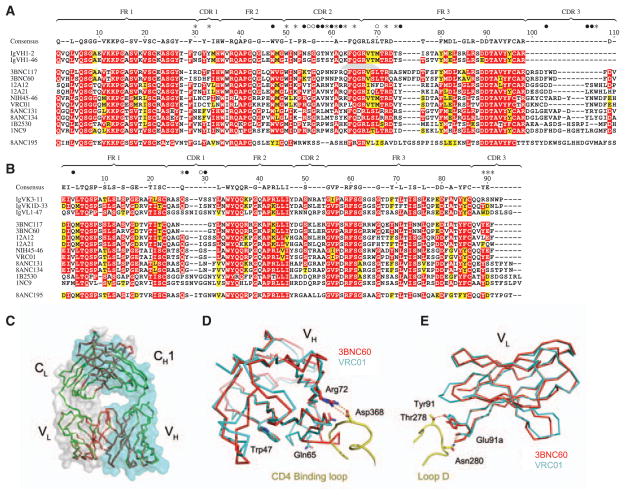
Passive transfer of broadly neutralizing HIV antibodies can prevent infection, which suggests that vaccines that elicit such antibodies would be protective. Thus far, however, few broadly neutralizing HIV antibodies that occur naturally have been characterized. To determine whether these antibodies are part of a larger group of related molecules, we cloned 576 new HIV antibodies from four unrelated individuals. All four individuals produced expanded clones of potent broadly neutralizing CD4-binding-site antibodies that mimic binding to CD4. Despite extensive hypermutation, the new antibodies shared a consensus sequence of 68 immunoglobulin H (IgH) chain amino acids and arise independently from two related IgH genes. Comparison of the crystal structure of one of the antibodies to the broadly neutralizing antibody VRC01 revealed conservation of the contacts to the HIV spike.


Figures

Fig. 1
Fig. 1
The 2CC core captures CD4bs antibodies. (A and B) Top line indicates bait used for sorting, and below is the patient number. The number in the center of the pies denotes the number of antibodies; slices are unique clones and are proportional to clone size. Membership of an antibody in a B cell clone is determined by sequence analysis, in particular CDR3s and shared V and J genes of paired heavy- and light-chain genes (table S3). (A) Pie charts show a summary of detected B cell clones irrespective of their binding epitopes. (★) Star indicates clones found in both YU2-gp140 and 2CC core–sorted cells. (B) Pie charts show the distribution of antibodies binding to CD4bs, CD4i, CD4ind. (equally affected by D368R and I420R), and core (not affected by either D368R or I420R). Dashed lines indicate antibodies that were cloned but could not be produced. (C) Representative ELISAs on YU2-gp120, mutants, and 2CC core.
Fig. 2
Fig. 2
Antibodies captured by 2CC core have broad and potent HIV-neutralizing activity. (A) HIV-neutralizing activity assayed on a limited panel of viruses. Top line indicates the donor number (table S2). Antibody clones are grouped, and individual clonal relatives are represented by a column grouped as in table S4. Each row represents one virus as indicated on the left. Colors indicate concentration of clones at the median inhibitory concentration (IC50): red, ≤0.1 μg/ml; orange, 0.1 to 1 μg/ml; yellow, 1 to 10 μg/ml; green, ≥10 μg/ml; and white, not neutralized at any concentration tested. (★) Star indicates the representatives selected for the extended virus panel (table S5). (B) HIV-neutralizing activity assayed on an extended panel of viruses (tables S5 and S6). (C) Neutralization summary graph comparing the published IC50 values of VRC01 (6) with NIH45-46 and 3BNC117 (tables S5 and S7). Each color represents a different HIV clade (black corresponds to CRF01AE, blue to clade B, green to clade G, pink to clade D, gray to clade CD, emerald to clade C, brown to clade ACD, magenta to clade AC, red to clade A, and orange to CRF02AG). Length of lines and size of circles are inversely proportional to IC50. The distance between the outer and inner circle, as well as from the inner circle to the center of the spiders, in each span is two logs in IC50 concentration.
Fig. 3
Fig. 3
Binding properties of antibodies. (A) Representative SPR sensor-grams for binding to YU2-gp140 and 2CC core by 12A12, 12A21 (Fig. 2B and table S5), and 12A germ line (GL)–reverted antibodies. (B) Graph shows KA for representative antibodies (tables S5 and S9). (C) Graph shows mean fluorescence intensity of CD4i antibody 3-67 (7) binding to Bal-expressing 293T cells after incubation with the indicated antibodies. Table indicates whether or not an antibody induces CD4i site accessibility (fig. S11).
Fig. 4
Fig. 4
Sequence and structural conservation of HAADs. (A) Amino acid alignment of 10 selected HAADs (table S5), their germline genes, and 8ANC195. Residues are numbered according to the 3BNC60 structure. Framework (FR) and CDR regions are indicated. Red shading shows amino acid identity; yellow shows biochemical similarity. The consensus is defined by 70% similarity between the 10 selected HAADs. The consensus sequence is shown above; dashes in this sequence indicate nonconserved residues. Contact residues between VRC01 and gp120 are shown above the consensus as closed circles for main-and side-chain interactions, open circles main chain only, and stars side chains only (5). (B) As in (A) for light chains. (C, D, and E) Crystal structure of 3BNC60 Fab. (C) Superimposed Cα traces of the two Fab molecules in the 3BNC60 asymmetric unit are shown in green and red. Semitransparent surfaces are used to outline the heavy (cyan) and light (gray) chains. (D) Superimposition of the 3BNC60 VH (red, Cα trace) and VRC01 VH (cyan, Cα trace) shown with a ribbon representation of the CD4-binding loop. The salt bridge between Arg71VRC01 and Asp368gp120 is shown as dashed lines. (E) Superimposition of the 3BNC60 VL (red, Cα trace). Hydrogen bonds between VRC01 and gp120 are shown as dashed lines.

Comment in

  • AIDS/HIV. Converging on an HIV vaccine.
    Korber B, Gnanakaran S. Korber B, et al. Science. 2011 Sep 16;333(6049):1589-90. doi: 10.1126/science.1211919. Science. 2011. PMID: 21921189 No abstract available.

References

    1. Walker LM, et al. PLoS Pathog. 2010;6:e1001028. - PMC - PubMed
    1. Doria-Rose NA, et al. J Virol. 2010;84:1631. - PMC - PubMed
    1. Gray ES, et al. J Virol. 2011;85:4828. - PMC - PubMed
    1. Mikell I, et al. PLoS Pathog. 2011;7:e1001251. - PMC - PubMed
    1. Zhou T, et al. Science. 2010;329:811. - PMC - PubMed

Publication types

MeSH terms