. 2011 Nov 1;39(20):8752-64.
doi: 10.1093/nar/gkr537. Epub 2011 Jul 15.

Resistance of bulky DNA lesions to nucleotide excision repair can result from extensive aromatic lesion-base stacking interactions

Affiliations

Resistance of bulky DNA lesions to nucleotide excision repair can result from extensive aromatic lesion-base stacking interactions

Dara A Reeves et al. Nucleic Acids Res. .

Abstract

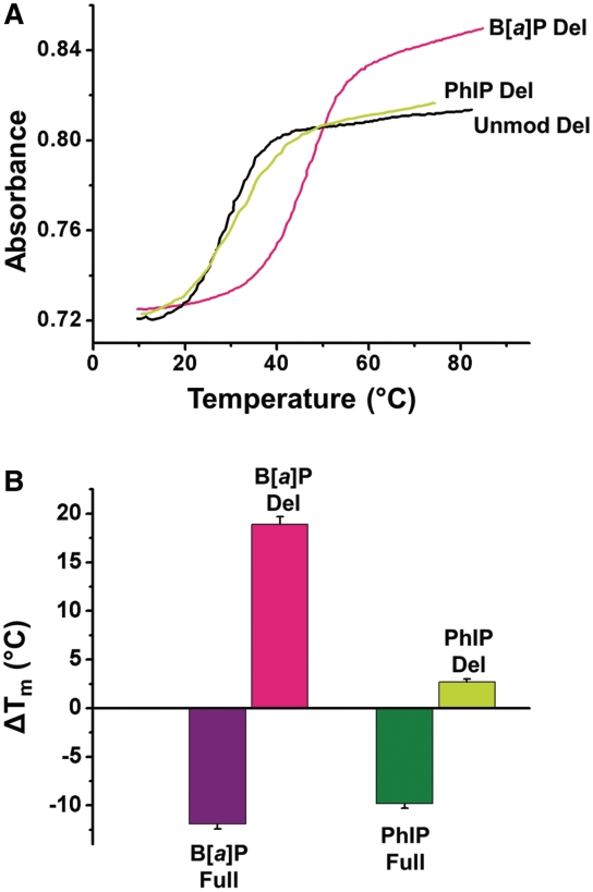
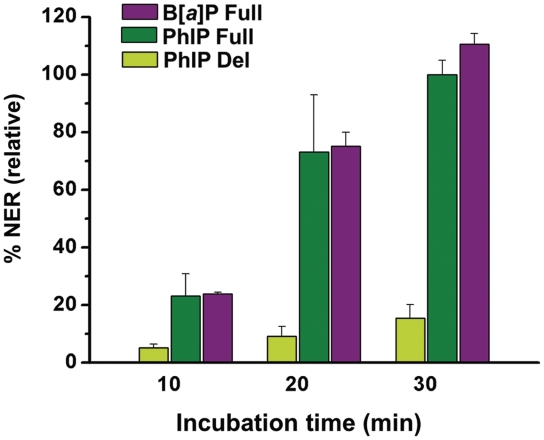
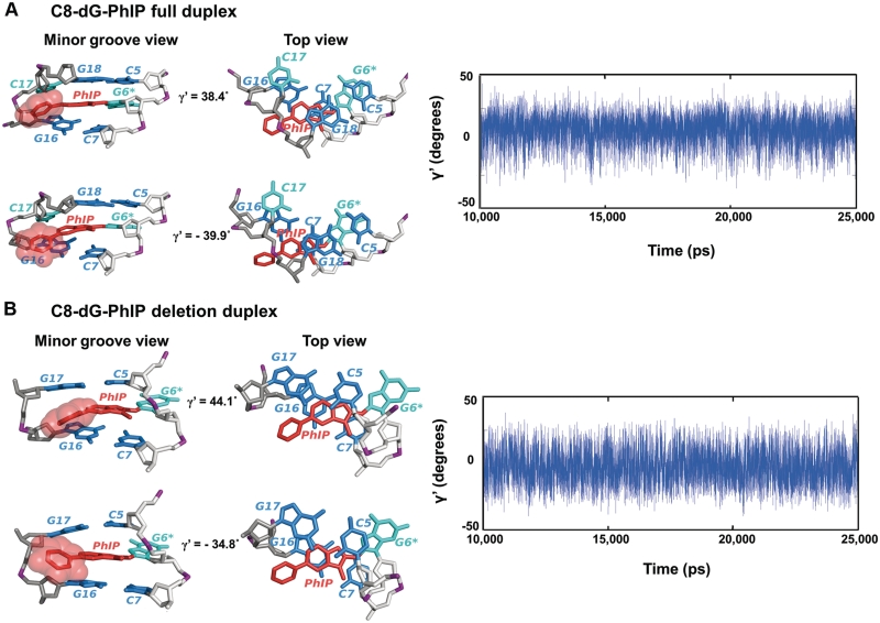
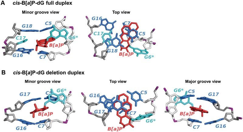
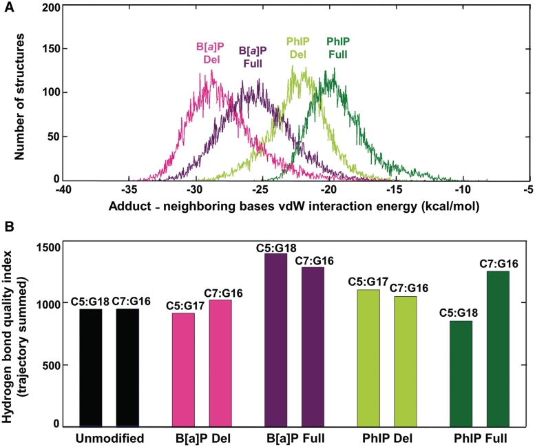
The molecular basis of resistance to nucleotide excision repair (NER) of certain bulky DNA lesions is poorly understood. To address this issue, we have studied NER in human HeLa cell extracts of two topologically distinct lesions, one derived from benzo[a]pyrene (10R-(+)-cis-anti-B[a]P-N(2)-dG), and one from the food mutagen 2-amino-1-methyl-6-phenylimidazo[4,5-b]pyridine (C8-dG-PhIP), embedded in either full or 'deletion' duplexes (the partner nucleotide opposite the lesion is missing). All lesions adopt base-displaced intercalated conformations. Both full duplexes are thermodynamically destabilized and are excellent substrates of NER. However, the identical 10R-(+)-cis-anti-B[a]P-N(2)-dG adduct in the deletion duplex dramatically enhances the thermal stability of this duplex, and is completely resistant to NER. Molecular dynamics simulations show that B[a]P lesion-induced distortion/destabilization is compensated by stabilizing aromatic ring system-base stacking interactions. In the C8-dG-PhIP-deletion duplex, the smaller size of the aromatic ring system and the mobile phenyl ring are less stabilizing and yield moderate NER efficiency. Thus, a partner nucleotide opposite the lesion is not an absolute requirement for the successful initiation of NER. Our observations are consistent with the hypothesis that carcinogen-base stacking interactions, which contribute to the local DNA stability, can prevent the successful insertion of an XPC β-hairpin into the duplex and the normal recruitment of other downstream NER factors.


Figures

Figure 1.
Figure 1.
Structures and sequences of cis-B[a]P-dG and C8-dG-PhIP-modified duplexes. (A) Chemical structure of the cis-B[a]P-dG adduct. The glycosidic torsion angle, χ for both modified duplexes is defined as O4′−C1′−N9−C4. The B[a]P-dG linkage site torsion angles, α′ and β′, are defined as follows: α′ = N1(G)−C2(G)−N2(G)−C10(B[a]P) and β′ = C2(G)−N2(G)−C10(B[a]P)−C9(B[a]P). (B) Chemical structure of the C8-dG-PhIP adduct. The PhIP-dG linkage site torsion angles, α′ and β′, are defined as follows: α′ = N9(G)−C8(G)−N(PhIP)−C2(PhIP) and β′ = C8(G)−N(PhIP)−C2(PhIP)−N1(PhIP). The torsion angle γ′ is defined as C7(PhIP)−C6(PhIP)−C1′(PhIP)−C2′(PhIP). (C) Sequence I. Sequence context of the 11/11-mer full duplex. (D) Sequence II. Sequence context of the 11/10-mer deletion duplex. G6*, colored in red, represents the lesion-modified guanine. (E) NMR solution structures for the cis-B[a]P-dG-modified 11/11mer full and 11/10mer deletion duplexes (34,35) and C8-dG-PhIP 11/11mer full duplex (33) utilized as initial structures for the MD simulations. The C8-dG-PhIP deletion duplex was modeled based on the cis-B[a]P-dG deletion duplex NMR structure (35), as described in ‘Materials and Methods’ section. The duplexes are viewed from the minor groove side. Hydrogen atoms and phosphate oxygen atoms are deleted for clarity. In the modified full duplexes, the lesion strand backbone is colored in light gray and the complementary strand is colored in dark gray. Phosphate atoms are colored purple. The adduct is colored red, the modified guanine and its partner base are colored cyan, and the neighboring bases are colored blue.
Figure 2.
Figure 2.
Thermal melting properties of cis-B[a]P-dG and C8-dG-PhIP-modified duplexes. (A) UV melting profiles of unmodified (Unmod), cis-B[a]P-dG (B[a]P) and C8-dG-PhIP (PhIP) modified deletion duplexes measured at 260 nm. The full (Full) and deletion (Del) duplexes are defined in Figure 1 and the Tm values and hyperchromicity values are summarized in Table 1. (B) Comparisons of the ΔTm = Tm (modified) – Tm (unmodified values) based on thermal melting data of Table 1. The melting points of the duplexes, Tm, are averages of 2–3 experiments.
Figure 3.
Figure 3.
NER assays in Human HeLa cell extracts. (A) Autoradiogram of a representative denaturing 12% polyacrylamide gel showing incisions of full (Full) and deletion (Del) 135-mer duplexes containing single cis-B[a]P-dG (B[a]P) and C8-dG-PhIP (PhIP) adducts in HeLa cell extracts at 10, 20 and 30 min of incubation. The zero time point samples serve as controls (these samples were incubated in heat-deactivated cell extracts, but otherwise underwent the same treatment). Lane Marker: oligonucleotide markers of different lengths. (B) Densitometry tracings of the 30 min lanes shown in the autoradiogram in Figure 3A. M: markers.
Figure 4.
Figure 4.
Incision kinetics of the internally labeled 135-mer modified duplexes in HeLa cell extracts. The incision efficiencies were normalized in each of the four independent experiments to the value obtained with the C8-dG-PhIP full duplex (relative value of 100) at the 30 min incubation time point. The averages and standard deviations shown were obtained from these normalized values. Note that the cis-B[a]P-dG deletion duplex is not incised.
Figure 5.
Figure 5.
Molecular dynamics of the C8-dG-PhIP-modified full and deletion duplexes. Hydrogen atoms and phosphate oxygen atoms are deleted for clarity. The lesion strand backbone is in light gray and the complementary strand is in dark gray. Phosphate atoms are purple. The adduct is red, the modified guanine and its partner base are cyan, and the neighboring bases are blue. (A) Dynamics of phenyl ring of the C8-dG-PhIP-modified full duplex. The structures with the highest and lowest γ′ values (38.4° and −39.9°) are shown on the left to emphasize the dynamics of the phenyl ring. The central trimer is viewed from the minor groove side (left) and along the helix axis (Top view, right). The time dependence of the γ′ torsion angle is shown on the right. (B) Dynamics of phenyl ring of the C8-dG-PhIP-modified deletion duplex. The structures with the highest and the lowest γ′ values (44.1° and −34.8°) are shown on the left to emphasize the dynamics of the phenyl ring. The central trimer is viewed from the minor groove side (left) and along the helix axis (Top view, right). The time dependence of the γ′ torsion angle is shown on the right.
Figure 6.
Figure 6.
Representative structures of the cis-B[a]P-dG-modified full and deletion duplexes. Hydrogen atoms and phosphate oxygen atoms are deleted for clarity. The lesion strand backbone is in light gray and the complementary strand is in dark gray. Phosphate atoms are purple. The adduct is red, the modified guanine and its partner base are cyan, and the neighboring bases are blue. (A) Best representative structure of the cis-B[a]P-dG-modified full duplex viewed from the minor groove and along the helix axis. The best representative structure viewed along the helix axis (Top view) is shown on the right to emphasize stacking interactions with neighboring bases. (B) Best representative structure of the cis-B[a]P-dG-modified deletion duplex viewed from the minor groove, along the helix axis and from the major groove. The best representative structure viewed along the helix axis (Top view) is shown in the middle to emphasize stacking interactions with neighboring bases. The best representative structure viewed from the major groove side is shown on the right to emphasize the wedge-like shape induced by the deletion.
Figure 7.
Figure 7.
Stabilizing and destabilizing properties of lesions in modified duplexes. (A) The population distribution of the vdW interaction energy between adduct aromatic rings and the neighboring bases. The vdW interaction energies were calculated as described in Supplementary Data. Energies for the cis-B[a]P-dG (B[a]P) and C8-dG-PhIP (PhIP) modified full duplexes (Full) are represented in dark green and purple, and in light green and magenta for the respective deletion duplexes (Del). Mean values and standard deviations (kcal/mol): B[a]P Del, −27.9 ± 2.6; B[a]P Full, −25.5 ± 2.6; PhIP Del, −22.4 ± 2.1; PhIP Full, −19.4 ± 2.2. (B) Trajectory summed hydrogen bond quality index for the C:G base pairs 5′ and 3′ to the lesion in the cis-B[a]P-dG (B[a]P) and C8-dG-PhIP (PhIP) modified deletion (Del) and full (Full) duplexes, and for the C:G base pairs at comparable positions in the unmodified duplex. The trajectory summed hydrogen bond quality indexes were calculated as described in the Supplementary Data. The specific base pairs are labeled at the top of the bars. The bars are color-coded as in Figure 7A.

Similar articles

Cited by

References

    1. Hanawalt PC, Spivak G. Transcription-coupled DNA repair: two decades of progress and surprises. Nat. Rev. Mol. Cell Biol. 2008;9:958–970. - PubMed
    1. Tornaletti S. DNA repair in mammalian cells: Transcription-coupled DNA repair: directing your effort where it's most needed. Cell Mol. Life Sci. 2009;66:1010–1020. - PMC - PubMed
    1. Gillet LC, Scharer OD. Molecular mechanisms of mammalian global genome nucleotide excision repair. Chem. Rev. 2006;106:253–276. - PubMed
    1. Mocquet V, Kropachev K, Kolbanovskiy M, Kolbanovskiy A, Tapias A, Cai Y, Broyde S, Geacintov NE, Egly JM. The human DNA repair factor XPC-HR23B distinguishes stereoisomeric benzo[a]pyrenyl-DNA lesions. EMBO J. 2007;26:2923–2932. - PMC - PubMed
    1. Mocquet V, Laine JP, Riedl T, Yajin Z, Lee MY, Egly JM. Sequential recruitment of the repair factors during NER: the role of XPG in initiating the resynthesis step. EMBO J. 2008;27:155–167. - PMC - PubMed

Publication types