. 2011 Oct;39(19):8404-15.
doi: 10.1093/nar/gkr555. Epub 2011 Jul 14.

A novel 4E-interacting protein in Leishmania is involved in stage-specific translation pathways

Affiliations

A novel 4E-interacting protein in Leishmania is involved in stage-specific translation pathways

Alexandra Zinoviev et al. Nucleic Acids Res. 2011 Oct.

Abstract

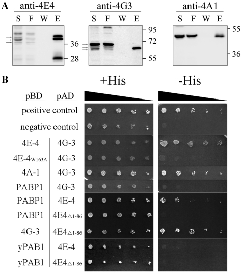
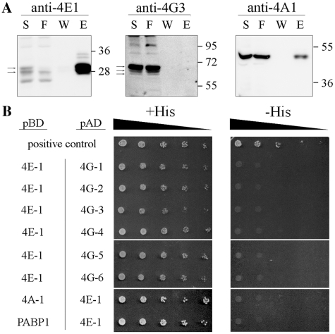
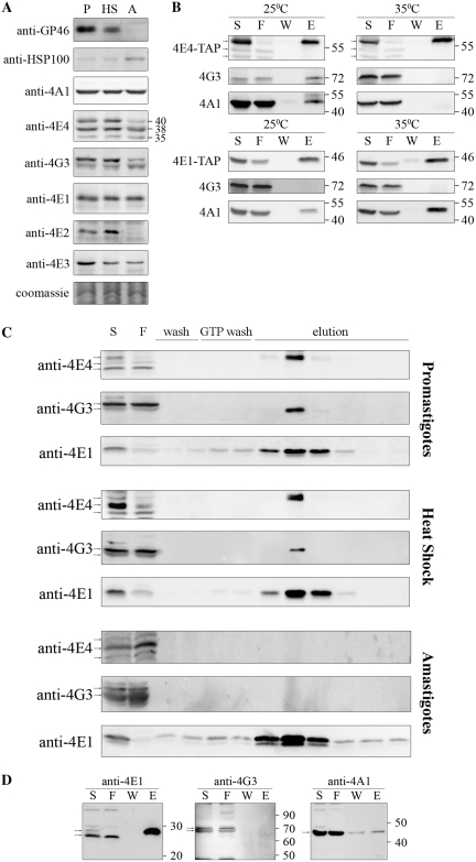
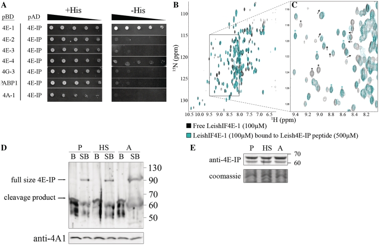
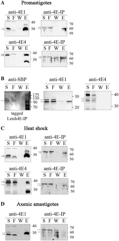
In eukaryotes, exposure to stress conditions causes a shift from cap-dependent to cap-independent translation. In trypanosomatids, environmental switches are the driving force of a developmental program of gene expression, but it is yet unclear how their translation machinery copes with their constantly changing environment. Trypanosomatids have a unique cap structure (cap-4) and encode four highly diverged paralogs of the cap-binding protein, eIF4E; none were found to genetically complement a yeast mutant failing to express eIF4E. Here we show that in promastigotes, a typical cap-binding complex is anchored through LeishIF4E-4, which associates with components of the cap-binding pre-initiation complex. In axenic amastigotes, expression of LeishIF4E-4 decreases and the protein does not bind the cap, whereas LeishIF4E-1 maintains its expression level and associates with the cap structure and with translation initiation factors. However, LeishIF4E-1 does not interact with eIF4G-like proteins in both life stages, excluding its involvement in cap-dependent translation. Using pull-down assays and mass-spectrometry, we identified a novel, non-conserved 4E-Interacting Protein (Leish4E-IP), which binds to LeishIF4E-1 in promastigotes, but not in amastigotes. Yeast two-hybrid and NMR spectroscopy confirmed the specificity of this interaction. We propose that Leish4E-IP is a translation regulator that is involved in switching between cap-dependent and alternative translation pathways.


Figures

Figure 1.
Figure 1.
Proteins associated with LeishIF4E-4 in promastigotes. (A) Pull-down experiments with the SBP-tagged LeishIF4E-4 from L. amazonensis promastigotes was done using affinity purification over streptavidin–Sepharose beads. Aliquots of the soluble extract (S, 1%), the flow-through (F, 1%), the final wash (W, 20%) and eluted proteins (E, 20%) were separated by SDS–PAGE (10–15%) and subjected to western blot analysis using specific antibodies against LeishIF4E-4, LeishIF4G-3 and LeishIF4A-1. Densitometric analysis that was normalized to the total protein amounts showed that the elution fraction contained 16, 9 and 3.4% of the total LeishIF4E-4, LeishIF4G-3 and LeishIF4A-1, respectively. The arrows indicate the specific reaction of the antibodies. (B) The yeast two-hybrid assay was performed by cotransfecting wild-type YRG-2 yeast strain with pBD fused to LeishIF4E-4, LeishIF4E-4 W163A mutant, LeishIF4A-1, LeishPABP-1, LeishIF4G-3 or yPAB1 and pAD-LeishIF4G-3, pAD-LeishIF4E-4 or pAD-LeishIF4E-4Δ1-86. Cells were cultured under restrictive (−His) and non-restrictive (+His) growth conditions, and spotted as described in ‘Materials and Methods’ section. Positive and negative controls were supplied by the manufacturer and expression of the tested proteins was verified by western blot analysis (Supplementary Figure S7).
Figure 2.
Figure 2.
Proteins associated with LeishIF4E-1 in promastigotes. (A) Pull-down experiments with the SBP-tagged LeishIF4E-1 from L. amazonensis promastigotes was done using affinity purification and further analysis as described in Figure 1A. The antibodies used were directed against LeishIF4E-1, LeishIF4G-3 and LeishIF4A-1. Densitometric analysis showed that the elution fraction contained 24, 0 and 3% of the total LeishIF4E-1, LeishIF4G-3 and LeishIF4A-1, respectively. (B) The yeast two-hybrid assay was performed by co-transfecting wild-type YRG-2 yeast strains with pBD fused to LeishIF4A-1, LeishPABP-1 or LeishIF4E-1 and with pAD fused to LeishIF4G-1 through -6 or to LeishIF4E-1. The cells were cultured and spotted as described in ‘Materials and Methods’ section. Expression of the tested proteins was verified by western blot analysis (Supplementary Figure S7).
Figure 3.
Figure 3.
Characterization of the LeishIF4F complex during stage differentiation. (A) Expression levels of LeishIF4F subunits in wild-type L. amazonensis, exposed or not to heat shock and differentiated in vitro. Cells were lysed and samples with 50 µg protein from the supernatants were resolved on SDS–PAGE (10–15%) and subjected to western blot analysis using specific antibodies against GP46, HSP100, LeishIF4E-1 to -4, LeishIF4G-3 and LeishIF4A-1. P, promastigotes from late log phase cultures; HS, late log promastigotes following a transient exposure to 33°C for 2 h; A, axenic amastigotes 9 days after differentiation. Coomassie staining of Tubulin was used to verify the protein loads. (B) Pull-down experiments with TAP tagged LeishIF4E-1 or LeishIF4E-4 from L. major promastigotes, exposed or not to heat shock (35°C, 2 h). Cell extracts were affinity purified over streptavidin-Sepharose beads and then over m7GTP-Sepharose column. The pulled-down proteins were analyzed as described in Figure 1A, using specific antibodies against LeishIF4E-1, LeishIF4E-4, LeishIF4G-3 and LeishIF4A-1. (C) m7GTP pulldown assays with wild-type L. amazonensis promastigotes, promastigotes after 2 h at 33°C and axenic-amastigotes, 9 days after differentiation. The cells were lyzed and loaded on a m7GTP-Sepharose column. The eluted proteins were analyzed as described in Figure 1A, using specific antibodies against LeishIF4E-1, LeishIF4E-4 and LeishIF4G-3. (D) Pull-down experiments in axenic-amastigotes of L. amazonensis expressing SBP tagged LeishIF4E-1, 9 days after differentiation. Proteins were purified and analyzed as described in Figure 1A, using specific antibodies against LeishIF4E-1, LeishIF4G-3 and LeishIF4A-1.
Figure 4.
Figure 4.
A novel 4E-IP homolog in Leishmania interacts with LeishIF4E-1 in vitro. (A) Yeast two-hybrid assay was performed by co-transfecting wild-type YRG-2 yeast strain with pBD fused to LeishIF4E-1 through -4, LeishIF4G-3, LeishPABP-1 and LeishIF4A-1 and pAD-Leish4E-IP. The cells were cultured and spotted as described in ‘Materials and Methods’ section. Expression of the tested proteins was verified by western blotting (Supplementary Figure S7). (B) Overlays [15N, 1H]TROSY-HSQC spectra of 15N-labeled LeishIF4E-1 (100 µM) bound to the human cap (m7GTP, 5 mM) without (black) and with (cyan) a non-labeled LeishIF4E-IP peptide (500 μM, VRTMYTREELLRIAT). In this figure, each peak corresponds to a specific LeishIF4E-1 amino acid. A region displaying chemical shift differences between LeishIF4E-1 unbound (black) and bound (cyan) to LeishIF4E-IP is boxed, and an enlargement is shown in (C). Examples of peaks that disappeared upon LeishIF4E-IP peptide binding are indicated by a black asterisk, while some remaining unaffected are indicated by a cyan asterisk. (D) Leish4E-IP appears to be easily cleaved in all life forms. Wild-type L. amazonensis promastigotes grown at normal temperatures (P), promastigotes after heat shock treatment at 33°C for 2 h (HS), and axenic amastigotes, 9 days after differentiation (A) were lyzed, washed and only the supernatants were used (B lanes); or lyzed by sample buffer without any prior treatment (SB lanes). The lysates were separated by SDS–PAGE (10–15%) and subjected to western blot analysis using specific antibodies against Leish4E-IP. Western blot analysis with anti-LeishIF4A-1 was used as a loading control. (E) Leish4E-IP is constitutively expressed in different Leishmania life forms. Wild-type L. amazonensis cells were treated and analyzed as described in Figure 3A, using specific antibody against Leish4E-IP. P, late log phase promastigotes; HS, late log promastigotes after 2 h of heat shock; A, axenic amastigotes, 9 days after differentiation.
Figure 5.
Figure 5.
Leish4E-IP interacts with LeishIF4E-1 and LeishIF4E-4 in promastigotes. Pull-down analysis with SBP tagged LeishIF4E-1, LeishIF4E-4 or Leish4E-IP from L. amazonensis was done using affinity purification and analysis as described in Figure 1A. Aliquots of the soluble extract (S, 1%), the flow-through (F, 1%), the final wash (W, 40% for A, C, D and 20% for B) and eluted proteins (E, 40% for A, C, D and 20% for B) were separated by SDS–PAGE (10–15%) and subjected to western blot analysis using specific antibodies against LeishIF4E-1, LeishIF4E-4, Leish4E-IP and SBP. The pull-down analysis was performed in promastigotes (A and B), promastigotes after 2 h at 33°C (C) and in axenic amastigotes, 9 days after differentiation (D) Densitometric analysis of panel A showed that the elution fraction contained 10.4 and 14% of LeishIF4E-1 and Leish4E-IP, respectively (top) and 8 and 12% of LeishIF4E-4 and Leish4E-IP, respectively (bottom). The wash fraction of Leish4E-IP that was pulled down by LeishIF4E-4 contained 7% of total protein, suggesting that the eluted fraction contained some protein that came down non-specifically.

Similar articles

Cited by

References

    1. Sonenberg N, Hinnebusch AG. Regulation of translation initiation in eukaryotes: mechanisms and biological targets. Cell. 2009;136:731–745. - PMC - PubMed
    1. Fletcher CM, McGuire AM, Gingras AC, Li H, Matsuo H, Sonenberg N, Wagner G. 4E binding proteins inhibit the translation factor eIF4E without folded structure. Biochemistry. 1998;37:9–15. - PubMed
    1. Fletcher CM, Wagner G. The interaction of eIF4E with 4E-BP1 is an induced fit to a completely disordered protein. Protein Sci. 1998;7:1639–1642. - PMC - PubMed
    1. Haghighat A, Mader S, Pause A, Sonenberg N. Repression of cap-dependent translation by 4E-binding protein 1: competition with p220 for binding to eukaryotic initiation factor-4E. EMBO J. 1995;14:5701–5709. - PMC - PubMed
    1. Mader S, Lee H, Pause A, Sonenberg N. The translation initiation factor eIF-4E binds to a common motif shared by the translation factor eIF-4 gamma and the translational repressors 4E-binding proteins. Mol. Cell. Biol. 1995;15:4990–4997. - PMC - PubMed

Publication types

MeSH terms

Substances