. 2011 Aug;134(Pt 8):2248-60.
doi: 10.1093/brain/awr161. Epub 2011 Jul 15.

The contribution of gliosis to diffusion tensor anisotropy and tractography following traumatic brain injury: validation in the rat using Fourier analysis of stained tissue sections

Affiliations

The contribution of gliosis to diffusion tensor anisotropy and tractography following traumatic brain injury: validation in the rat using Fourier analysis of stained tissue sections

Matthew D Budde et al. Brain. 2011 Aug.

Abstract

Diffusion tensor imaging is highly sensitive to the microstructural integrity of the brain and has uncovered significant abnormalities following traumatic brain injury not appreciated through other methods. It is hoped that this increased sensitivity will aid in the detection and prognostication in patients with traumatic injury. However, the pathological substrates of such changes are poorly understood. Specifically, decreases in fractional anisotropy derived from diffusion tensor imaging are consistent with axonal injury, myelin injury or both in white matter fibres. In contrast, in both humans and animal models, increases in fractional anisotropy have been suggested to reflect axonal regeneration and plasticity, but the direct histological evidence for such changes remains tenuous. We developed a method to quantify the anisotropy of stained histological sections using Fourier analysis, and applied the method to a rat controlled cortical impact model to identify the specific pathological features that give rise to the diffusion tensor imaging changes in subacute to chronic traumatic brain injury. A multiple linear regression was performed to relate the histological measurements to the measured diffusion tensor changes. The results show that anisotropy was significantly increased (P < 0.001) in the perilesioned cortex following injury. Cortical anisotropy was independently associated (standardized β = 0.62, P = 0.04) with the coherent organization of reactive astrocytes (i.e. gliosis) and was not attributed to axons. By comparison, a decrease in white matter anisotropy (P < 0.001) was significantly related to demyelination (β = 0.75, P = 0.0015) and to a lesser extent, axonal degeneration (β = -0.48, P = 0.043). Gliosis within the lesioned cortex also influenced diffusion tensor tractography, highlighting the fact that spurious tracts in the injured brain may not necessarily reflect continuous axons and may instead depict glial scarring. The current study demonstrates a novel method to relate pathology to diffusion tensor imaging findings, elucidates the underlying mechanisms of anisotropy changes following traumatic brain injury and significantly impacts the clinical interpretation of diffusion tensor imaging findings in the injured brain.


Figures

Figure 1
Figure 1
In vivo DTI following controlled cortical impact in the rat brain. (A) A region of increased fractional anisotropy (FA) is observed in the perilesion cortex with its orientation maintained perpendicular to the cortical surface. (B) Regions of interest were placed on the T2-weighted images in the perilesion, contralateral and control cortices and white matter for quantification. (C) In five controlled cortical impact and five naïve control animals, the significant increase in perilesion cortical fractional anisotropy was the result of an increase in axial (parallel) diffusivity with no change in radial (perpendicular) diffusivity. In the white matter, the decreased perilesion fractional anisotropy was a result of an increase in radial diffusivity with no change in axial diffusivity. Values indicate mean ± SD (n = 5 for each). *P < 0.05. CC = corpus callosum; CCI = controlled cortical impact.
Figure 2
Figure 2
Quantification of anisotropy in histological sections. (A) A brain section stained with DiI is shown from a control animal at the region of the corpus callosum. The white boxes indicate examples from regions of low (B) and high (C) expected anisotropy. (B and C) Each image subsection was windowed to reduce edge effects and the 2D Fourier transform (2DFT) was applied. Radial integration and principal component analysis was applied to derive anisotropy (Aer) and orientation (θ). (D) The calculated orientation and anisotropy were displayed as hue and saturation, respectively, and the 2D ellipse was overlaid in white.
Figure 3
Figure 3
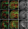
Comparison of ex vivo DTI and whole-brain DiI sections. Ex vivo DTI maps colour-coded for anisotropy and orientation are shown for four slices from a control brain (left). The similarly coloured maps computed from the corresponding histological sections using the proposed method (middle) are similar in appearance to the DTI maps. The original DiI-stained sections are shown for reference (right), with the numbers indicating the anatomical location in reference to bregma.
Figure 4
Figure 4
Cellular components of controlled cortical impact lesions using immunofluorescence and anisotropy maps. Within the injured controlled cortical impact cortex and white matter, glial fibrillary acidic protein (GFAP)-positive astrocytes are prolific. In the cortex, lesser changes in MAP2, SMI31 and SMI32 were evident, with all four stains displaying increased anisotropy as shown in the Aer maps (insets). In the white matter, increased SMI32 staining and decreased myelin basic protein (MBP) structural integrity were evident in both the stained sections and the resulting Aer maps (insets).
Figure 5
Figure 5
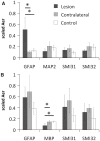
Quantitative histology results. (A) In the cortex, glial fibrillary acidic protein (GFAP)-derived anisotropy was significantly greater in the perilesioned tissue compared with the contralateral and control cortices. (B) In the white matter, myelin basic protein (MBP) anisotropy was significantly decreased in the perilesioned tissue compared with the contralateral and control white matter. Values indicate mean ± SD (n = 5 for each). *P < 0.05.
Figure 6
Figure 6
Whole-brain in vivo DTI tractography. (A) DTI tractography of normal rat brain (left) and the controlled cortical impact (CCI) brain (right) reveals numerous tracts propagating into the controlled cortical impact lesion (arrow). (B) Regions of interest were segmented using boundaries that included the cortical surface (green), the medial aspect of the lateral ventricle and the posterior edge of the corpus callosum. (C) The region of interest mask applied to the tractography results demonstrates numerous tracts that course through both the cortical lesion and the corpus callosum. (D) Among all animals, the mean tract length of cortical tracts is significantly greater in the lesioned cortex compared with either the contralateral or control brain.
Figure 7
Figure 7
Comparison of diffusion tensor tractography and histology-derived tractography. Three representative controlled cortical impact animals are shown. (A) Ex vivo 3D diffusion tensor tractography (DTT) maps depict tracts propagating in and along the lesion periphery similar to those observed in vivo. (B) 2D diffusion tensor tractography was subsequently performed to allow direct comparison to the 2D histology-derived tractography. (C) 2D tractography maps from the glial fibrillary acidic protein (GFAP)-stained sections revealed similarities to the DTI-derived maps near the lesion border. (D) The coherent orientation of astrocytes is shown on confocal images from selected regions. (E) 2D tractography maps from the SMI32-stained sections revealed fewer, if any, tracts propagating into the cortex near the lesion periphery. (F) A loss of structural integrity is noted for both the injured white matter and grey matter along the lesion border.

References

    1. Anderova M, Vorisek I, Pivonkova H, Benesova J, Vargova L, Cicanic M, et al. Cell death/proliferation and alterations in glial morphology contribute to changes in diffusivity in the rat hippocampus after hypoxia-ischemia. J Cereb Blood Flow Metab. 2010;31:894–907. - PMC - PubMed
    1. Arfanakis K, Haughton VM, Carew JD, Rogers BP, Dempsey RJ, Meyerand ME. Diffusion tensor MR imaging in diffuse axonal injury. AJNR Am J Neuroradiol. 2002;23:794–802. - PMC - PubMed
    1. Armitage PA, Bastin ME. Selecting an appropriate anisotropy index for displaying diffusion tensor imaging data with improved contrast and sensitivity. Magn Reson Med. 2000;44:117–21. - PubMed
    1. Armitage PA, Bastin ME, Marshall I, Wardlaw JM, Cannon J. Diffusion anisotropy measurements in ischaemic stroke of the human brain. MAGMA. 1998;6:28–36. - PubMed
    1. Axer M, Amunts K, Grässel D, Palm C, Dammers J, Axer H, et al. A novel approach to the human connectome: ultra-high resolution mapping of fiber tracts in the brain. NeuroImage. 2011;54:1091–101. - PubMed

Publication types

MeSH terms