. 2011 Jul 15;25(14):1486-98.
doi: 10.1101/gad.2059211.

Dynamic transcriptional events in embryonic stem cells mediated by the super elongation complex (SEC)

Affiliations

Dynamic transcriptional events in embryonic stem cells mediated by the super elongation complex (SEC)

Chengqi Lin et al. Genes Dev. .

Abstract

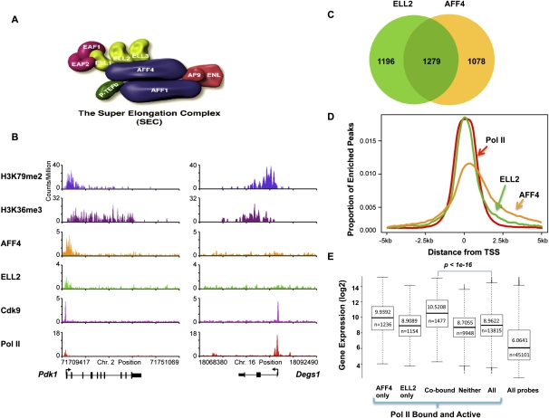
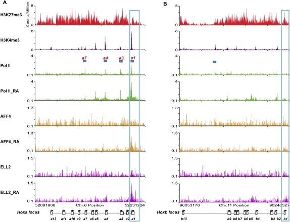
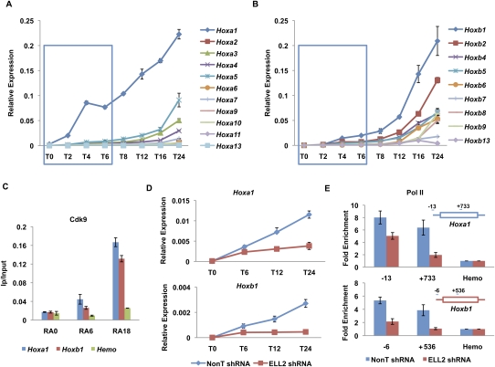
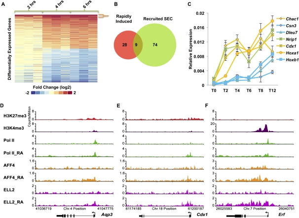
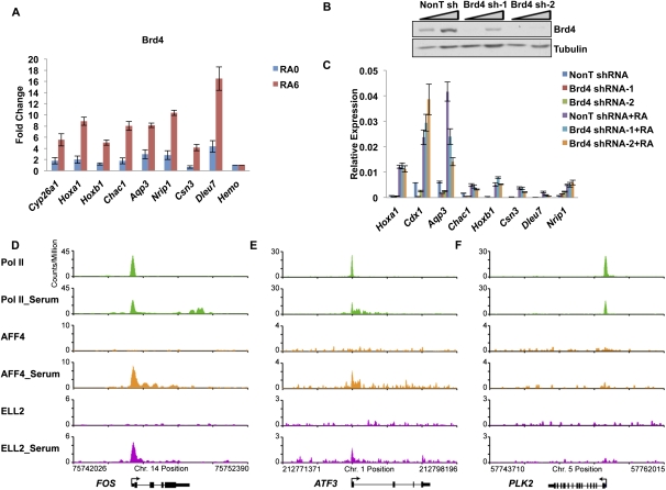
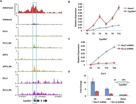
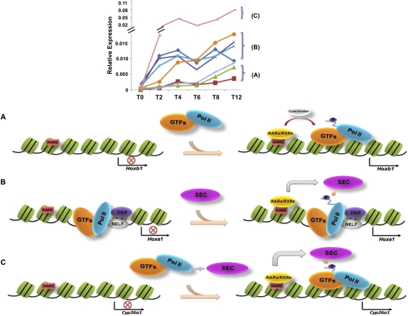
Transcriptional regulation of developmentally controlled genes is at the heart of differentiation and organogenesis. In this study, we performed global genomic analyses in murine embryonic stem (ES) cells and in human cells in response to activation signals. We identified an essential role for the ELL (eleven-nineteen lysine-rich leukemia gene)/P-TEFb (positive transcription elongation factor)-containing super elongation complex (SEC) in the regulation of gene expression, including several genes bearing paused RNA polymerase II (Pol II). Paused Pol II has been proposed to be associated with loci that respond rapidly to environmental stimuli. However, our studies in ES cells also identified a requirement for SEC at genes without paused Pol II, which also respond dynamically to differentiation signals. Our findings suggest that SEC is a major class of active P-TEFb-containing complexes required for transcriptional activation in response to environmental cues such as differentiation signals.


Figures

Figure 1.
Figure 1.
Global occupancy of the SEC subunits in mouse ES cells. (A) Schematic representation of the SEC. The SEC is a P-TEFb-containing complex that contains various combinations of four types of proteins: ELL1–3; EAF1–2; AFF1 and AFF4; and AF9 and ENL. P-TEFb itself consists of Cdk9 and CycT1/2 and is best characterized as an RNA Pol II CTD kinase. (B) Genome-wide analysis of SEC components AFF4, ELL2, and Cdk9 by ChIP-seq in ES cells found SEC enriched at a subset of actively transcribed genes. Shown are two genes with high levels of expression in ES cells. (Left panel) The Pdk1 gene is occupied by the SEC subunits ELL2 and AFF4. (Right panel) The Degs1 gene does not have significant levels of the SEC components AFF4 and ELL2. H3K36 trimethylation (H3K36me3) and H3K79 dimethylation (H3K79me2) data from Marson et al. (2008) are shown as markers of actively transcribed genes. (C) Venn diagram analysis of AFF4- and ELL2-occupied genes. Around 50% of AFF4-enriched genes are also occupied by ELL2, demonstrating that in mouse ES cells, these two proteins share a similar global occupancy. (D) Histogram of the genome-wide occupancy of AFF4, ELL2, and Pol II. The canonical TSS of each gene in the genome was used to measure the distance to the nearest bound region, which is plotted if falling within 5 kb of the TSS. This analysis shows that SEC components are enriched over the TSS, similar to Pol II occupancy. (E) AFF4 and ELL2 co-occupy highly transcribed genes. The dark lines in the box plots and the number above the line indicate the median level of expression for the gene subset indicated. The number below the line indicates the number of Affymetrix probe sets that correspond to the gene subset. Probe sets for ELL2 and AFF4 cobound genes show significantly higher expression compared with all Pol II-bound and active genes (P < 1 × 10⁻¹⁶ by Wilcoxon two-sample rank sum test). The gene subset containing neither AFF4 nor ELL2 also shows some highly expressed genes. Genes were called active if they were determined present on the array by the MAS5 algorithm.
Figure 2.
Figure 2.
The Hoxa1 promoter is preloaded with Pol II and recruits SEC after RA treatment in ES cells. (A) Bivalent marks, paused Pol II, and SEC recruitment to the Hoxa cluster. In ES cells, the whole Hoxa cluster is highly enriched for H3K27me3, and also contains H3K4me3 at the promoters of a subset of genes, including Hoxa1, Hoxa3, Hoxa4, and Hoxa7. These regions are preloaded with Pol II (bars indicate regions that have both a bivalent mark and Pol II). (B) Bivalent marks and paused Pol II are both largely absent from the Hoxb genes, which do not recruit SEC after 6 h of RA treatment. While H3K27me3 marks the whole cluster of Hoxb genes, only Hoxb4, Hoxb7, and Hoxb9 contain H3K4me3 at their promoters and can be considered bivalent. There is no significant Pol II detected on the promoters of the Hoxb genes in ES cells. The bar marks a peak of significant Pol II that does not correspond to a known gene feature. Before RA treatment, there is no detectable AFF4 and ELL2 signal on the Hoxa or Hoxb cluster genes. Both AFF4 and ELL2 are recruited to the Hoxa1, but not the Hoxb1, gene promoter after exposure to RA for 6 h. Blue boxes highlight the Hoxa1 and Hoxb1 genes. Expanded views of the Hoxa1 and Hoxb1 regions are shown in Supplemental Figure S4.
Figure 3.
Figure 3.
SEC is required for the rapid induction of the Hoxa1 gene. (A,B) RT-qPCR analysis of Hoxa and Hoxb cluster genes upon RA treatment. ES cells were treated with RA for different time points as indicated. Total RNAs were extracted from these cells and then subjected to RT-qPCR analysis using an Applied Biosystems' custom TaqMan array card. Hoxa1 was the first Hox gene to be induced by RA. Compared with Hoxa1, the induction of Hoxb1 was much slower within the first 6 h of RA treatment. The blue boxes indicate the first three RA induction time points. (C) Cdk9 is recruited to both the Hoxa1 and Hoxb1 gene promoters. Cdk9 ChIP was performed to measure its enrichment on Hoxa1 and Hoxb1 after RA treatment. A hemoglobin gene, Hba (Hemo), serves as a nontranscribed control gene. (D) ELL2 RNAi inhibits the induction of Hoxa1 and Hoxb1 by RA. shRNA targeting ELL2 or nontargeting shRNA (NonT) was introduced by lentiviral infection for 3 d before RA treatment. (E) Knockdown of ELL2 reduces Pol II occupancy at Hoxa1 and Hoxb1 after 6 h of RA treatment. Pol II occupancy was assayed by ChIP at the start site of transcription and in the ORF of Hoxa1 and Hoxb1 in RA-induced cells. Pol II is reduced in the ORF of both Hoxa1 and Hoxb1, and Hoxb1 also shows dramatically reduced levels of Pol II at its promoter after ELL2 RNAi. The Hoxa1 promoter, but not the Hoxb1 promoter, has prebound Pol II before RA treatment (see Fig. 2; Supplemental Fig. S4). Error bars represent the standard deviation.
Figure 4.
Figure 4.
SEC regulates the rapid induction of RA signaling. (A, left panel) Microarray analyses of RA induction of ES cells as a function of time (2, 4, and 6 h) in biological triplicate. Differentially expressed probes (twofold or more) at the sixth hour post-induction compared with no induction are shown. Thirty-seven genes were induced twofold or more at each of the 2-, 4-, and 6-h time points (demarcated by the orange bracket). (B) Of the 37 induced genes, nine of them recruited SEC (ELL2 and AFF4). Newly recruited SEC genes are cobound at 6 h post-induction and are not cobound before induction. (C) RT-qPCR analysis of some of the induced genes identified from the microarray analysis. ES cells were treated with RA for the indicated time points, 0 h (T0), 2 h (T2), 4 h (T4), 6 h (T6), 8 h (T8), and 12 h (T12). Genes that recruit SEC are shown in yellow and genes that do not recruit SEC are shown in blue. Nrip1, which does not recruit SEC but is rapidly induced, is shown in green. Error bars represent the standard deviation. (D–F) Examples of ChIP-seq data showing SEC recruitment to RA-induced genes. Shown are Aqp3, Cdx1, and Erf, three of the nine genes from B.
Figure 5.
Figure 5.
Brd4 is broadly present, but not broadly required, for RA induction of genes. (A) ChIP of Brd4 at RA-6-induced genes. Brd4 levels significantly increase at all RA-6-induced genes tested. The Hba gene serves as a nontranscribed control gene. (B) shRNA-mediated knockdown of Brd4. Two different shRNA constructs targeting Brd4 and a nontargeting shRNA (NonT) were introduced by lentiviral infection for 3 d before RA treatment. Brd4 levels were significantly reduced by Western analysis. Triangles indicate titrations of cell extracts. Tubulin serves as a loading control. (C) Induction of genes with RA is not broadly affected by Brd4 knockdown. Several genes, identified in Figure 4 as rapidly induced, were assayed for expression levels before and after RA treatment. Only Aqp3 showed a significant decrease in its induction. Error bars represent the standard deviation. (D–F) SEC is recruited to the immediate early genes in HCT-116 cells after serum stimulation, genes previously identified as regulated by Brd4-containing P-TEFb complexes. HCT-116 cells were starved for 40 h before the 30-min add-back of serum. (D–F) Genome browser track files of three serum-induced genes, which were previously shown to be rapidly induced after serum stimulation (Donner et al. 2010). SEC is recruited to ATF3 and FOS concomitant with release of paused Pol II into the gene body. PLK2 is shown for comparison as a gene induced by serum that does not recruit SEC.
Figure 6.
Figure 6.
The rapid induction of Cyp26a1 does not involve preloaded Pol II. (A) Pol II, H3K4me3, and H3K27me3 occupancy analysis of the Cyp26a1 gene before RA induction. Before RA treatment, the Cyp26a1 promoter is significantly enriched for H3K27me3, with lower levels of H3K4me3. However, there is no detectable Pol II on the promoter. AFF4, ELL2, and Pol II are newly recruited to the Cyp26a1 gene promoter upon RA treatment. (B) RT-qPCR analysis of Cyp26a1 mRNA levels upon RA treatment. ES cells were treated with RA for the indicated time points, 0 h (T0), 2 h (T2), 4 h (T4), 6 h (T6), 8 h (T8), and 12 h (T12). Total RNAs were extracted from these treated cell samples and then subjected to RT-qPCR analysis. (C) ELL2 RNAi inhibits the induction of Cyp26a1 by RA. shRNA targeting ELL2 or a nontargeting shRNA (NonT) was introduced by lentiviral infection for 3 d before RA treatment. (D) Knockdown of ELL2 reduces Pol II occupancy at Cyp26a1 after 24 h RA treatment. The Hba gene serves as a nontranscribed control gene. Error bars represent the standard deviation.
Figure 7.
Figure 7.
Diverse mechanisms for rapid activation of genes during development. The top panel shows that rapidly activated genes can be further subdivided into distinct categories, A–C. (A) The Hoxb1 gene newly recruits Pol II and GTFs in a classical gene activation mechanism, where RAR/RXR binds in the presence of RA and, with the help of coactivators, recruits GTFs and Pol II. (B) Paused Pol II, with DSIF/NELF, is present at the TSS of developmentally regulated genes, such as Hoxa1. In the presence of RA, RAR/RXR recruits SEC to stimulate transcription elongation through phosphorylation of the DSIF/NELF and the Pol II CTD. (C) Cyp26a1, a developmentally regulated gene that lacks paused Pol II, is induced by RA in a SEC-dependent manner. All of the same factors are present after RA treatment as seen at Hoxa1, but Cyp26a1 is induced to higher levels, suggesting that paused Pol II may serve to help regulate activation to equivalent levels.

References

    1. Abu-Abed S, Dolle P, Metzger D, Beckett B, Chambon P, Petkovich M 2001. The retinoic acid-metabolizing enzyme, CYP26A1, is essential for normal hindbrain patterning, vertebral identity, and development of posterior structures. Genes Dev 15: 226–240 - PMC - PubMed
    1. Alexander T, Nolte C, Krumlauf R 2009. Hox genes and segmentation of the hindbrain and axial skeleton. Annu Rev Cell Dev Biol 25: 431–456 - PubMed
    1. Bernstein BE, Mikkelsen TS, Xie X, Kamal M, Huebert DJ, Cuff J, Fry B, Meissner A, Wernig M, Plath K, et al. 2006. A bivalent chromatin structure marks key developmental genes in embryonic stem cells. Cell 125: 315–326 - PubMed
    1. Boettiger AN, Levine M 2009. Synchronous and stochastic patterns of gene activation in the Drosophila embryo. Science 325: 471–473 - PMC - PubMed
    1. Bres V, Yoh SM, Jones KA 2008. The multi-tasking P-TEFb complex. Curr Opin Cell Biol 20: 334–340 - PMC - PubMed

Publication types

MeSH terms

Substances