. 2011 Sep;10(9):1207-18.
doi: 10.1128/EC.05089-11. Epub 2011 Jul 15.

Vacuolar protein sorting protein 13A, TtVPS13A, localizes to the tetrahymena thermophila phagosome membrane and is required for efficient phagocytosis

Affiliations

Vacuolar protein sorting protein 13A, TtVPS13A, localizes to the tetrahymena thermophila phagosome membrane and is required for efficient phagocytosis

Haresha S Samaranayake et al. Eukaryot Cell. 2011 Sep.

Abstract

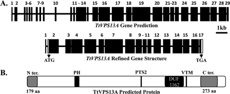
Vacuolar protein sorting 13 (VPS13) proteins have been studied in a number of organisms, and mutations in VPS13 genes have been implicated in two human genetic disorders, but the function of these proteins is poorly understood. The TtVPS13A protein was previously identified in a mass spectrometry analysis of the Tetrahymena thermophila phagosome proteome (M. E. Jacobs et al., Eukaryot. Cell 5:1990-2000, 2006), suggesting that it is involved in phagocytosis. In this study, we analyzed the structure of the macronuclear TtVPS13A gene, which was found to be composed of 17 exons spanning 12.5 kb and was predicted to encode a protein of 3,475 amino acids (aa). A strain expressing a TtVPS13A-green fluorescent protein (GFP) fusion protein was constructed, and the protein was found to associate with the phagosome membrane during the entire cycle of phagocytosis. In addition, Tetrahymena cells with a TtVPS13A knockout mutation displayed impaired phagocytosis. Specifically, they grew slowly under conditions where phagocytosis is essential, they formed few phagosomes, and the digestion of phagosomal contents was delayed compared to wild-type cells. Overall, these results provide evidence that the TtVPS13A protein is required for efficient phagocytosis.


Figures

Fig. 1.
Fig. 1.
The Tetrahymena TtVPS13A gene and encoded protein. (A) Comparison of the TtVPS13A gene prediction (TGD accession number 8.m00549) and the refined gene structure based on RT-PCR analysis. Exons are denoted by black boxes and numbered; introns and 5′- and 3′-untranslated regions are shown by black horizontal lines. Translation start and stop sites are also indicated. (B) Diagram of the predicted 3,475-aa TtVPS13A protein. N- and C-terminal regions that are well conserved among VPS13 proteins are indicated as gray boxes, along with putative DUF1162 and pleckstrin homology (PH) domains, a second peroxisomal signal (PTS2), and a vacuole-targeting motif (VTM).
Fig. 2.
Fig. 2.
Construction of a Tetrahymena strain expressing a TtVPS13A::GFP fusion protein. (A) Diagrams of the TtVPS13A::GFP construct, the 3′ end of the endogenous macronuclear TtVPS13A locus, and the expected integration product following transformation. Coding regions are shown as rectangles, and the 3′-UTR and flanking regions are shown as lines. The arrowheads represent the positions of the PCR primers used to determine if there was a complete replacement of endogenous copies by the construct. Diagrams are not drawn to scale. (B) PCR analysis to assess gene replacement in TtVPS13A::GFP transformants. A portion of an agarose gel is shown, with PCR products obtained from total genomic DNAs of four assorted subclones (lanes 1 to 4) as well as wild-type CU428.2 cells (lane WT), using oligonucleotides CH3ENDF, GFPF, and CHUTRXHO1R as primers. The positions of selected size markers (in kilobase pairs) and the PCR products expected for the GFP-tagged (GFP) (322 bp) and endogenous TtVPS13A (ENDO.) (246 bp) gene copies are indicated. (C) Western blot analysis of total cell proteins from transformant TtVPS13A::GFP1 cells (lane 1), TtVPS13A::GFP2 cells (lane 2), the wild-type CU428.2 cell line (lane 3), and a Tetrahymena cell line overexpressing a RAB7::GFP fusion protein (lane 4), using an affinity-purified anti-GFP polyclonal antibody (1:2,000 dilution). The expected positions of the full-length TtVPS13A::GFP (VPS13A) and RAB7::GFP (Rab7) proteins are indicated on the right, and the sizes and positions of selected protein molecular mass markers (lane M) are indicated on the left in kilodaltons.
Fig. 3.
Fig. 3.
Subcellular localization of the TtVPS13A::GFPA5:10 fusion protein. (A) Confocal images of fixed Tetrahymena TtVPS13A:GFPA5:10 and CU428.2 cells that had been either fed with nonfluorescent latex beads for 30 min or grown in the absence of latex beads. For slide preparation, cells were collected by centrifugation and resuspended in SlowFade antifade agent. Fluorescent and bright-field (BF) images of cells are shown. Thin arrows indicate selected bead-filled phagosomes, and arrowheads indicate selected small vesicles. (B) Time course analysis of the TtVPS13A::GFP association with phagosomes. Cells were fed latex beads for 10 min and examined by confocal fluorescence (GFP) and bright-field microscopy over the next 120 min. These results were reproducible in multiple, independent time course analyses.
Fig. 4.
Fig. 4.
Timing of molecular events during phagocytosis. (A) Phagosome acidification was assessed by the preincubation of cells with LysoTracker dye for 10 min, followed by feeding with latex beads for 2 min. Red fluorescence confocal (FLUOR.) and differential interference contrast (DIC) microscopy images of cells analyzed over the next 2-h time period are shown. Selected acidified phagosomes displaying bright fluorescence are indicated by arrows. (B) The delivery of cathepsin B to phagosomes was analyzed by feeding latex beads to a cell line overexpressing a cathepsin B::GFP fusion protein (19). GFP fluorescence and DIC images collected at 1, 5, 30, 60, and 120 min are shown. (C) Graph of the percentage of VPS13A-containing phagosomes (diamonds), cathepsin B-associated phagosomes (▵), and acidified phagosomes (○) versus time.
Fig. 5.
Fig. 5.
Generation of a TtVPS13A gene knockout. (A) Diagrams of the knockout construct (KO), which contains a neomycin resistance gene (NEO4R) under the control of the cadmium-inducible MTT1 promoter (open boxes), and the 5′ end of the endogenous TtVPS13A gene (exons are shown as black boxes). The integration of the construct by homologous recombination results in the deletion of parts of intron 2 and exon 3. Arrows denote the primers used in PCR and RT-PCR analyses to confirm that the transformants carried the expected deletion. The TtVPS13AΔ transformants are expected to have the NEO4 gene between exon 2 and exon 3. The arrows indicate the locations of the primers used in the PCR and RT-PCR analyses. (B) PCR analysis to determine the ratio of wild-type (WT) and deleted (knockout) gene copies in the transformants before and after phenotypic assortment. The 1% agarose gel shown contains the PCR mixtures from the TtVPS13AA4 (A4) and TtVPS13AB2 (B2) transformants, wild-type (WT) CU428.2 cells, and the assorted subclones TtVPS13AA4PA (A4PA) and TtVPS13AB2PA (B2PA). PCR was carried out on total genomic DNA from transformants and the WT by using the pNEO4MTT1 and CHIN2R reverse primers, along with forward primer CHIN2F (see above). The positions of the expected WT (0.22 kb) and knockout mutant (0.40 kb) PCR products are indicated. (C) Gel with RT-PCR products representing TtVPS13A and GRL1 gene transcripts in wild-type cells and the TtVPS13AA4PA and TtVPS13AB2PA knockout subclones. RT-PCR was carried out by using forward primer CH1617L and reverse primer CHEX3R to amplify TtVPS13A transcripts, and forward primer EX5L and reverse primer rEX2 were included to amplify GRL1 transcripts, which served as an internal control. The sizes of the expected PCR products for TtVPS13A and GRL1 are indicated in kilobase pairs.
Fig. 6.
Fig. 6.
Characterization of growth and phagocytosis in TtVPS13A knockout cell lines. (A and B) Growth curves (log of cells/ml versus time) of Tetrahymena CU428.2 (square symbols) and the TtVPS13AA4PA (triangles) and TtVPS13AB2PA (circles) knockout subclones in EPP medium (A) and under conditions requiring phagocytosis (Tris buffer supplemented with Klebsiella bacteria) (B) are shown. Similar results were obtained with two additional repeats of each analysis. (C) Graph showing the number of phagosomes per cell in wild-type CU428.2 cells (squares), TtVPS13AA4PA (triangles), and TtVPS13AB2PA (circles) versus time after the addition of 2-μm red fluorescent latex beads. Data shown are averages of data from two experiments that yielded similar results.
Fig. 7.
Fig. 7.
Digestion of GFP-labeled Bacillus spores. (A) Paired DIC and fluorescence micrographs of wild-type CU428.2 (left panels) and mutant TtVPS13AA4PA cells (right panels) at various times (20 to 120 min) following the addition of decoated Bacillus subtilis spores expressing a GFP-labeled protein. Arrows denote selected brightly fluorescent phagosomes that contain intact spores. (B) Plot of the percentage of phagosomes with intact spores versus time for CU428.2 cells (squares) and the TtVPS13AA4PA transformant (triangles). Mean values derived from three experiments are shown, with bars representing the standard errors of the means.

Similar articles

Cited by

References

    1. Allen L. A., Aderem A. 1996. Mechanisms of phagocytosis. Curr. Opin. Immunol. 8:36–40 - PubMed
    1. Allen R. D., Fok A. K. 2000. Membrane trafficking and processing in Paramecium. Int. Rev. Cytol. 198:277–318 - PubMed
    1. Allen R. D., Wolf R. W. 1979. Membrane recycling at the cytoproct of Tetrahymena. J. Cell Sci. 35:217–227 - PubMed
    1. Bowen S., et al. 2007. The phagocytic capacity of neurons. Eur. J. Neurosci. 25:2947–2955 - PubMed
    1. Brickner J. H., Fuller R. S. 1997. SOI1 encodes a novel, conserved protein that promotes TGN-endosomal cycling of Kex2p and other membrane proteins by modulating the function of two TGN localization signals. J. Cell Biol. 139:23–36 - PMC - PubMed

Publication types

MeSH terms

LinkOut - more resources